. 2007 Jan;19(1):131-47.
doi: 10.1105/tpc.106.040782. Epub 2007 Jan 19.

Interactions among PIN-FORMED and P-glycoprotein auxin transporters in Arabidopsis

Affiliations

Interactions among PIN-FORMED and P-glycoprotein auxin transporters in Arabidopsis

Joshua J Blakeslee et al. Plant Cell. 2007 Jan.

Abstract

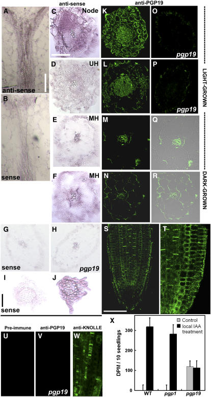
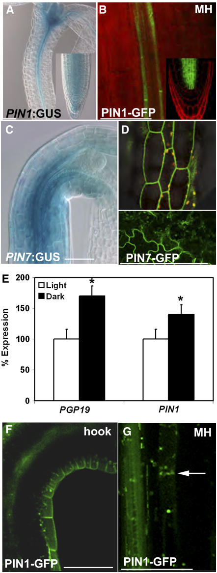
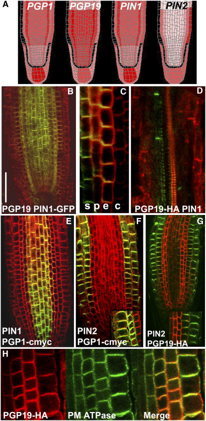
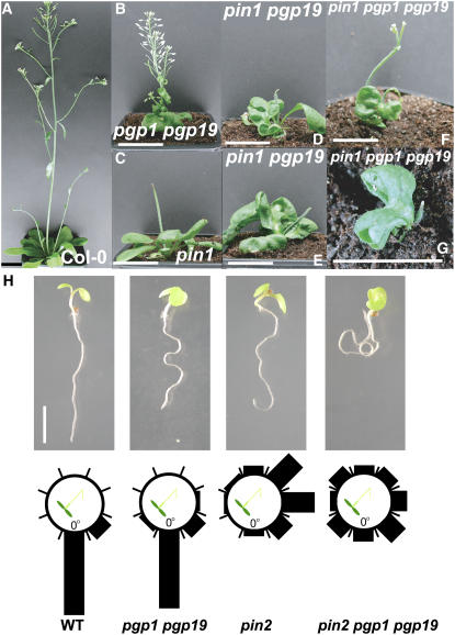
Directional transport of the phytohormone auxin is established primarily at the point of cellular efflux and is required for the establishment and maintenance of plant polarity. Studies in whole plants and heterologous systems indicate that PIN-FORMED (PIN) and P-glycoprotein (PGP) transport proteins mediate the cellular efflux of natural and synthetic auxins. However, aromatic anion transport resulting from PGP and PIN expression in nonplant systems was also found to lack the high level of substrate specificity seen in planta. Furthermore, previous reports that PGP19 stabilizes PIN1 on the plasma membrane suggested that PIN-PGP interactions might regulate polar auxin efflux. Here, we show that PGP1 and PGP19 colocalized with PIN1 in the shoot apex in Arabidopsis thaliana and with PIN1 and PIN2 in root tissues. Specific PGP-PIN interactions were seen in yeast two-hybrid and coimmunoprecipitation assays. PIN-PGP interactions appeared to enhance transport activity and, to a greater extent, substrate/inhibitor specificities when coexpressed in heterologous systems. By contrast, no interactions between PGPs and the AUXIN1 influx carrier were observed. Phenotypes of pin and pgp mutants suggest discrete functional roles in auxin transport, but pin pgp mutants exhibited phenotypes that are both additive and synergistic. These results suggest that PINs and PGPs characterize coordinated, independent auxin transport mechanisms but also function interactively in a tissue-specific manner.


Figures

Figure 1.
Figure 1.
PGP19 Localization in Light- and Dark-Grown Seedlings. (A) to (J) Localization of PGP19 expression using in situ hybridization in 7-d-old seedlings. All seedlings are wild type (Wassilewskija [Ws]) unless indicated otherwise. Bars = 50 μm. (A), (C) to (F), (H), and (J) Antisense probe. (B), (G), and (I) Sense probe. (A) to (E) and (G) to (J) Light-grown seedlings. (F) Dark-grown seedling. (A) Cotyledonary node and upper hypocotyl longitudinal section with an antisense probe shows signal in the vascular bundle. (B) Cotyledonary node and upper hypocotyl longitudinal section with a sense probe shows no signal. (C) Cotyledonary node cross section shows strong signal throughout the node. (D) Upper hypocotyl (UH; below the cotyledonary node) cross section shows signal throughout the hypocotyl. (E) Mid hypocotyl (MH) cross section shows signal restricted to the vascular bundle. (F) Mid hypocotyl cross section of a dark-grown seedling shows signal throughout the hypocotyl. (G) Hypocotyl cross section with a sense probe shows no signal. (H) Hypocotyl cross section of a light-grown pgp19 seedling with an antisense probe shows no signal. (I) Root cross section with a sense probe shows no signal. (J) Root cross section with an antisense probe shows strong signal throughout the root. (K) to (U) Immunohistochemical localization of PGP19 using PGP19-specific antiserum, unless indicated otherwise. All are 7-d-old wild-type (Ws) seedlings unless indicated otherwise. Bars = 50 μm. (K) to (Q) and (S) to (W) Light-grown seedlings. (N) and (R) Dark-grown seedlings. (K) Cotyledonary node cross section shows strong signal throughout tissue. (L) Upper hypocotyl (below the cotyledonary node) cross section shows signal throughout tissue. (M) Mid hypocotyl cross section shows signal in the vascular bundle. (N) Mid hypocotyl cross section of a dark-grown seedling shows signal throughout tissue. (O) Cotyledonary node cross section of a pgp19 seedling does not show signal. (P) Hypocotyl cross section of a pgp19 seedling does not show signal. (Q) Bright-field overlay of (M). (R) Bright-field overlay of (N). (S) Whole mount root tip of a 5-d-old light-grown seedling shows signal in the stele, endodermis, pericycle, and cortex. (T) Detail of the root tip shown in (S). (U) Preimmune serum in whole mount root tip of a 5-d-old seedling does not show signal. (V) Whole mount root tip of a 5-d-old pgp19 seedling does not show signal. (W) KNOLLE, using anti-KNOLLE (green), signal localizes at the cell plate during cytokinesis in a 5-d-old seedling. (X) Movement of [3H]IAA from stelar flow into the cortical/epidermal apoplast of mature root tissues before (gray bars) and after (black bars) application of additional cold IAA to the root–shoot transition zone. [3H]IAA was initially applied to the shoot apex in a discontinuous system to establish polar flow. Data are means ± sd (n = 10).
Figure 2.
Figure 2.
PIN1 Localization in Light- and Dark-Grown Seedlings. (A) ProPIN1:GUS activity is observed at the shoot apex, in vascular tissue, and in the root tip of a light-grown seedling. (B) ProPIN1:PIN1-GFP fluorescence is observed in the xylem parenchyma in hypocotyl and root tip in a light-grown seedling. MH, mid hypocotyl. (C) ProPIN7:PIN7-GUS activity is observed at the node and throughout the hypocotyl in a dark-grown seedling. The abundance of PIN7 in leaf cells may contribute to the substrate specificity seen in protoplast transport assays (Geisler et al., 2005). (D) ProPIN7:PIN7-GFP fluorescence is apolar in the epidermis of hypocotyls (top) and cotyledons (bottom). (E) Expression of PGP19 and PIN1 in 5-d-old light- and dark-grown seedlings. The light-grown value was set to 100% for each gene. Data are means ± sd (n = 3). * P < 0.05. (F) ProPIN1:PIN1-GFP fluorescence is observed in the xylem parenchyma and epidermis in the apical hook of a dark-grown seedling. (G) ProPIN1:PIN1-GFP fluorescence is observed in the xylem parenchyma and adjacent cortical cell (arrow) in the hypocotyl of a dark-grown seedling. Bars = 100 μm.
Figure 3.
Figure 3.
Overlapping Patterns of Expression and Localization. (A) Heat map of root tips from the AREX database (www.arexdb.org) showing the expression of PGP1, PGP19, PIN1, and PIN2. Relative expression is proportional to color intensity. (B) to (H) PGP localizations are shown in red; PIN and ATPase localizations are shown in green; colocalization is shown in yellow. All images show coimmunolocalizations in 5-d-old light-grown seedlings. Bar = 50 μm. (B) PGP19 and ProPIN1:PIN1-GFP in the root. (C) ProPGP19:PGP19-HA and PIN1 in the root. s, stele; p, pericycle; e, endodermis; c, cortex. ProPGP19:PGP19-HA complements the phenotype of pgp19. The PGP19-HA construct was used previously by Petrášek et al. (2006). (D) ProPGP19:PGP19-HA and PIN1 in the hypocotyl. (E) ProPGP1:PGP1-cmyc and PIN1 in the root. (F) ProPGP1:PGP1-cmyc and PIN2 in the root. (G) ProPGP19:PGP19-HA and PIN2 in the root. (H) ProPGP19:PGP19-HA and plasma membrane (PM) ATPase in the root.
Figure 4.
Figure 4.
Phenotypes of pgp pin Mutants. (A) Wild-type Columbia (Col-0) plant. (B) pgp1 pgp19 plant. (C) pin1 plant. (D) pin1 pgp19 plant (E) Another pin1 pgp19 plant. (F) pin1 pgp1 pgp19 plant. (G) Another pin1 pgp1 pgp19 plant. (H) Root phenotypes of the wild type, pgp1 pgp19, pin2, and pin2 pgp1 pgp19. The number of roots in 30° sectors of a circle were counted and expressed as a percentage of the total number of roots. Vertical position represents normal gravitropic response. Values were calculated using 40 seedlings per experiment. Bar = 5 cm in (A) to (G) and 1 cm in (H).
Figure 5.
Figure 5.
Substrate Specificity in Planta. [3H]BA transport in wild-type (Ws), pgp1, pgp19, and pin1 seedlings. Data are means ± sd and are expressed as percentages of wild-type values (n = 3). * P < 0.05.
Figure 6.
Figure 6.
Protein–Protein Interactions Are Indicated in Coimmunoprecipitation and Yeast Two-Hybrid Assays. (A) to (C) Coimmunoprecipitation assays. (A) Detergent-solubilized proteins from 5-mg microsomal membranes from the wild type (Ws) immunoprecipitated (IP) with PGP1/19 antiserum (which strongly binds PGP1 but PGP19 less strongly; left), PGP19 antiserum (middle), or AHA2 antibody (right) as a control. The blots were probed with PIN1 antibody. PGP1/19, PGP19, and PIN1 coimmunoprecipitated. Samples were run on 12% gels. (B) Detergent-solubilized proteins from 10-mg microsomal membranes from Pro35S:PGP19-HA transformants coimmunoprecipitated with PIN1 antiserum (left). In the reciprocal experiment, PGP19 coimmunoprecipitated with PIN1-GFP in ProPIN1:PIN1-GFP transformants (right). Samples were run on 8% gels. (C) Detergent-solubilized proteins from 10-mg microsomal membranes from ProPGP1:PGP1-cmyc transformants immunoprecipitated with cmyc antibody and probed with either PIN1 (left) or AHA2 (right) antiserum as a control. PGP1-cmyc coimmunoprecipitated PIN1 but not AHA2. A nonspecific band is observed on both blots. Coimmunoprecipitations are not quantitative. Samples were run on 8% gels. (D) Yeast two-hybrid assays. Soluble loops of PIN1, PIN2, and the C terminus of PGP19 were used in yeast two-hybrid interaction assays and growth and α-galactosidase assays for MEL1 reporter gene expression. Empty binding domain (BD) or activation domain (AD) vectors were transformed with PGP19-AD vector or PGP19-BD vector, respectively, as negative controls. AD and BD constructs for reverse assays were also analyzed, and the results were the same as the data presented. In addition, the PIN-AD and PIN-BD assays showed no growth or α-galactosidase activity. Three transformants from 10 independent transformations for each pair of constructs were analyzed. Values shown are means ± sd (n = 2). * P < 0.001, as determined by Student's t test.
Figure 7.
Figure 7.
Coexpression of PIN and PGP Transporters Increases Substrate Specificity, Inhibitor Sensitivity, and Efflux. (A) to (F) Efflux of radiolabeled substrates from HeLa cells expressing PGP1, PGP19, PIN1, or PIN2. Data are means with sum sd (n = 3). (A) Net efflux of [3H]IAA, [3H]IAA in the presence of NPA, or [3H]BA in HeLa cells expressing PGP1, PGP19, PIN1, PIN2, and AUX1. Baselines from a new set of experiments are presented only for the purpose of comparison with coexpression studies. Data for PGP1 and PIN2 were originally published by Geisler et al. (2005) and Petrášek et al. (2006). Efflux of substrates by PGP1 and PGP19 were significantly different from empty vector values (P < 0.05), and NPA inhibition of IAA efflux by PGP19 was significantly different compared with IAA alone (P < 0.05). BA efflux by PIN1 was significantly different from empty vector values (P < 0.05), as was IAA efflux by PIN2 (P = 0.05). (B) [3H]1-NAA export by PGP1, PGP4, PGP19, PIN1, and PIN2. PIN1 specificity for 1-NAA was not different from IAA. PGPs and PIN2 had less affinity for 1-NAA than for IAA. (C) Net efflux of [3H]IAA, [3H]IAA in the presence of NPA, or [3H]BA in HeLa cells coexpressing PIN1 with PGP1 or PGP19. IAA efflux by PIN1+PGP1 or PGP19 was significantly different from that of each protein alone (P < 0.05). NPA inhibition of IAA efflux by PIN1+PGP1 or PGP19 was significantly different compared with IAA alone (P < 0.05). BA efflux was not different from empty vector values (P > 0.05). (D) Net efflux of [3H]IAA, [3H]IAA in the presence of NPA, or [3H]BA in HeLa cells coexpressing PIN2 with PGP1 or PGP19. IAA efflux by PIN2+PGP1 or PGP19 was significantly different from that of each protein alone (P < 0.05). NPA inhibition of IAA efflux by PIN2+PGP1 or PGP19 was significantly different compared with IAA alone (P < 0.05). BA efflux was not different from empty vector values (P > 0.05). (E) Net efflux of [3H]IAA in HeLa cells coexpressing PGP4 with PIN1 or PIN2. Coexpression of PGP4 with PIN1 reversed PGP4-mediated influx, resulting in auxin efflux. Coexpression of PGP4 with PIN2 led to a synergistic increase in auxin influx. IAA efflux by PGP4 was significantly different from that of empty vector alone (P < 0.05). IAA efflux by PGP4+PIN1 or PIN2 was significantly different compared with each protein alone (P < 0.05). (F) Net efflux of [3H]IAA in HeLa cells expressing AUX1, PGP1, or PGP4. AUX1 expressed in HeLa cells mediated IAA influx. When AUX1 was coexpressed with PGP4, an additive effect on net IAA influx was observed. When AUX1 was coexpressed with PGP1, net IAA transport was not observed. (G) and (H) Efflux of radiolabeled IAA and BA from yeast cells expressing PGP1, PIN1, or PIN2. Data are means with se (n = 5 for IAA and n = 3 for BA). (G) Net [3H]IAA export in yeast cells expressing PIN1, PIN2, or PGP1 or coexpressing PGP1 with PIN1 or PIN2. Coexpression of PGP1 with PIN1 synergistically increased auxin efflux, whereas coexpression of PGP1 with PIN2/AGR1/EIR1 led to decreased auxin efflux. (H) Net [14C]BA export in yeast cells expressing PIN1, PIN2, or PGP1 or coexpressing PGP1 with PIN1 or PIN2. Cells coexpressing PGP1 with PIN1 or PIN2 exhibited reduced BA efflux.

References

    1. Ambudkar, S.V., Sauna, Z.E., Gottesman, M.M., and Szakacs, G. (2005). A novel way to spread drug resistance in tumor cells: Functional intercellular transfer of P-glycoprotein (ABCB1). Trends Pharmacol. Sci. 26 385–387. - PMC - PubMed
    1. Benkova, E., Michniewicz, M., Sauer, M., Teichmann, T., Seifertova, D., Jurgens, G., and Friml, J. (2003). Local, efflux-dependent auxin gradients as a common module for plant organ formation. Cell 115 591–602. - PubMed
    1. Bennett, M.J., Marchant, A., Green, H.G., May, S.T., Ward, S.P., Millner, P.A., Walker, A.R., Schulz, B., and Feldmann, K.A. (1996). Arabidopsis AUX1 gene: A permease-like regulator of root gravitropism. Science 273 948–950. - PubMed
    1. Birnbaum, K., Shasha, D.E., Wang, J.Y., Jung, J.W., Lambert, G.M., Galbraith, D.W., and Benfey, P.N. (2003). A gene expression map of the Arabidopsis root. Science 302 1956–1960. - PubMed
    1. Blakeslee, J.J., Bandyopadhyay, A., Peer, W.A., Makam, S.N., and Murphy, A.S. (2004). Relocalization of the PIN1 auxin efflux facilitator plays a role in phototropic responses. Plant Physiol. 134 28–31. - PMC - PubMed

Publication types

MeSH terms

Substances