. 2007 Feb 1;178(3):1791-9.
doi: 10.4049/jimmunol.178.3.1791.

Regulatory T cell vaccination without autoantigen protects against experimental autoimmune encephalomyelitis

Affiliations

Regulatory T cell vaccination without autoantigen protects against experimental autoimmune encephalomyelitis

Javier Ochoa-Repáraz et al. J Immunol. .

Abstract

Regulatory T (T(reg)) cells show promise for treating autoimmune diseases, but their induction to elevated potency has been problematic when the most optimally derived cells are from diseased animals. To circumvent reliance on autoantigen-reactive T(reg) cells, stimulation to myelin-independent Ags may offer a viable alternative while maintaining potency to treat experimental autoimmune encephalomyelitis (EAE). The experimental Salmonella vaccine expressing colonization factor Ag I possesses anti-inflammatory properties and, when applied therapeutically, reduces further development of EAE in SJL mice. To ascertain T(reg) cell dependency, a kinetic analysis was performed showing increased levels of FoxP3(+)CD25(+)CD4(+) T cells. Inactivation of these T(reg) cells resulted in loss of protection. Adoptive transfer of the vaccine-induced T(reg) cells protected mice against EAE with greater potency than naive or Salmonella vector-induced T(reg) cells, and cytokine analysis revealed enhanced production of TGF-beta, not IL-10. The development of these T(reg) cells in conjunction with immune deviation by Th2 cells optimally induced protective T(reg) cells when compared those induced in the absence of Th2 cells. These data show that T(reg) cells can be induced to high potency to non-disease-inducing Ags using a bacterial vaccine.


Conflict of interest statement

Disclosures

The authors have no financial conflict of interest.

Figures

FIGURE 1.
FIGURE 1.
Oral Salmonella-CFA/I (H696) treatment reduced EAE clinical scores in PLP139–151-challenged SJL/J mice (A) and decreased spinal cord demyelination (B) and inflammation (B and C). A, Mice therapeutically treated with H696 showed a significant reduction in their clinical scores with all mice recovering from EAE. Mice treated with Salmonella vector (strain H647) also recovered, but presented greater clinical scores when compared with PBS-treated mice. Depicted are the combined results from four separate experiments for a total of 20 mice/group: *, p < 0.001 for PBS vs H647 and PBS vs H696. B, H696-treated group showed minimal demyelination (LFB) and inflammatory (H&E stain) cell infiltration (indicated by arrows) at 14 days postchallenge when compared with PBS-dosed group; the H647-treated mice showed increased demyelination and inflammation. Representative samples from six mice are depicted. C, FACS analysis of spinal cord leukocytes was performed 14 days after challenge to assess the types of inflammatory cells: neutrophils, CD11b+SK208+; macrophages, CD11b+SK208F4/80+; and T cells, TCRβ+. Minimal leukocytes were detected in H696-treated mice unlike that observed for PBS- or H647-treated mice; representative samples of five mice per group are depicted.
FIGURE 2.
FIGURE 2.
Elevated Th2-type cytokines contribute to the therapeutic treatment by H696 vaccine by splenic (A) and CLN (B) lymphocytes with reductions in IFN-γ. Splenic (A) and CLN (B) lymphocytes were collected 14 days after EAE challenge (8 days after oral Salmonella vaccination) and cultured in the presence of OVA, purified CFA/I fimbriae, PLP139–151, or medium. Data depict the mean of three different experiments ± SEM. *, p < 0.001 for H696 vs PBS and H696 vs H647.
FIGURE 3.
FIGURE 3.
Oral Salmonella vaccination enhances the percentage of Treg cells expressing FoxP3 in both the mucosal and systemic compartments. A, CLN lymphocytes taken from SJL mice 14 days after EAE challenge (treated with H696, H647, or PBS 6 days postchallenge) and were examined by FACS for the presence of CD25+CD4+ T cells. Each group showed stimulation of Treg cells, but mice treated with H647 or H696 had increased numbers, and these were >90% FoxP3+. B, A kinetic analysis was performed on spleen, MLN, PP, SMLN, and deep CLN. Increases in FoxP3+ Treg cells were observed for Salmonella-treated groups. *, p < 0.001 for PBS vs H647 and PBS vs H696. C and D, Oral vaccination with H696 only (no PLP139–151 challenge) results in FoxP3+ Treg cells in spleen, MLN, PP, SMLN, and (C and D) CLN peaking ~2 wk after immunization (mean of five mice per group ± SEM).
FIGURE 4.
FIGURE 4.
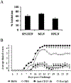
The Treg cells induced by Salmonella-CFA/I vaccination suppress effector T cells, and their in vivo inactivation results in enhanced clinical disease. A, CD25CD4+ and CD25+CD4+ T cells from spleens, MLN, and HNLN were cell sorted from mice orally immunized with H696 for 2 wk. Teff (1 × 10⁵) cells with feeder (1 × 10⁵) cells were cultured with CFA/I fimbriae and with or without Treg (5 × 10⁴) cells for 4 days. During the last 18 h of culture, cells were pulsed with [3H]TdR. Proliferation in the absence of Treg cells was set at zero inhibition. Depicted is the percent inhibition upon coculture with splenic, MLN, or HNLN Treg cells. One of three experiments is depicted. B, One week before PLP139–151 challenge, mice (five per group) orally immunized with H696 were treated in vivo with anti-CD25 mAb, rat IgG, or PBS on days −5 and −2 before challenge. Inactivation of Treg cells provoked greater EAE than the PBS-treated group. Normal rat IgG-treated mice vaccinated with H696 showed minimal EAE. *, p < 0.001 for anti-CD25-treated group vs H696-vaccinated or H696-vaccinated plus normal rat IgG-treated mice.
FIGURE 5.
FIGURE 5.
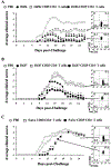
Adoptive transfer of Treg cells obtained from Salmonella-CFA/I-immunized mice confers greater protection against EAE challenge than Salmonella vector-induced Treg cells whereas those from naive mice failed to protect; protection is TGF-β dependent. Immune CD25+CD4+ T cells and CD25CD4+ T cells from 2-wk H696 (A)- and H647 (B)-immunized SJL mice were obtained from pooled spleens, MLN, and HNLN, purified by cell sorting, and 6 × 10⁵ Treg or Teff cells were injected i.v. into naive SJL mice. PBS-, H696-, and H647-immunized mice were used as negative and positive controls, respectively. Recipient mice given immune H696 Treg cells conferred total protection against EAE, whereas mice given Teff cells showed only partial protection with greater clinical disease than H696-vaccinated mice. *, p < 0.001 for PBS vs Treg cell group, CD25CD4+ T cell group, or H696-vaccinated group. Treg cells obtained from H647-vaccinated mice were less protective, and Teff cells failed to protect against EAE. C, Naive SJL Treg and Teff cells were purified by cell sorting of lymphocytes from MLN, HNLN, and spleens. Treg or CD25CD4+ T cells (6 × 10⁵) were injected i.v. into naive recipients 1 day before PLP139–151 challenge. While adoptive transfer of naive Treg cells delayed EAE onset, these failed to protect against EAE development. *, p < 0.001 for PBS vs naive Treg cell group or naive CD25CD4+ T cell group.
FIGURE 6.
FIGURE 6.
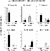
Oral vaccination with Salmonella-CFAH elicits TGF-β-producing Treg cells and IL-4-, IL-10-, and IL-13-producing Teff cells in contrast to IL-17-producing, but no Th2-type cytokine-producing, Teff cells upon vaccination with Salmonella vector. Cell-sorted CD25+CD4+ and CD25CD4+ T cells from mice orally immunized with H696 or H647 were evaluated for cytokine production following anti-CD3 and anti-CD28 costimulation. Treg cells had reduced IFN-γ production but elevated TGF-β when compared with Teff cells. IL-4, IL-10, and IL-13 segregated with the Teff cells induced by vaccination with H696. Teff cells from H647-vaccinated mice did not produce any Th2-type cytokines but rather produced elevated levels of IFN-γ. H647-induced Treg cells did produce TGF-β (although less than H696-vaccinated mice) and IL-17. Thus, protection conferred by Salmonella-CFA/I-induced Treg cells is because of reduced IFN-γ production and increased TGF-β, as well as in part supported by immune deviation by the Teff cells. *, p < 0.001 represents differences in cytokine production between CD25+CD4+ and CD25CD4+ T cells, and †, p < 0.001 represents differences in cytokine production between H696- and H647-sorted cells.

Similar articles

Cited by

References

    1. Sospedra M, and Martin R. 2005. Immunology of multiple sclerosis. Annu. Rev. Immunol 23: 683–747. - PubMed
    1. Steinman L 1996. Multiple sclerosis: a coordinated immunological attack against myelin in the central nervous system. Cell 85: 299–302. - PubMed
    1. Zeine R, Heath D, and Owens T. 1993. Enhanced response to antigen within lymph nodes of SJL/J mice that were protected against experimental allergic encephalomyelitis by T cell vaccination. J. Neuroimmunol 44: 85–94. - PubMed
    1. Shaw MK, Kim C, Hao HW, Chen F, and Tse HY. 1996. Induction of myelin basic protein-specific experimental autoimmune encephalomyelitis in C57BL/6 mice: mapping of T cell epitopes and T cell receptor Vβ gene segment usage. J. Neurosci. Res 45: 690–699. - PubMed
    1. Greer JM, Sobel RA, Sette A, Southwood S, Lees MB, and Kuchroo VK. 1996. Immunogenic and encephalitogenic epitope clusters of myelin proteolipid protein. J. Immunol 156: 371–379. - PubMed

Publication types

MeSH terms