. 2007 Mar 23;282(12):9150-61.
doi: 10.1074/jbc.M609304200. Epub 2007 Jan 21.

Pyrimethamine as a potential pharmacological chaperone for late-onset forms of GM2 gangliosidosis

Affiliations

Pyrimethamine as a potential pharmacological chaperone for late-onset forms of GM2 gangliosidosis

Gustavo H B Maegawa et al. J Biol Chem. .

Abstract

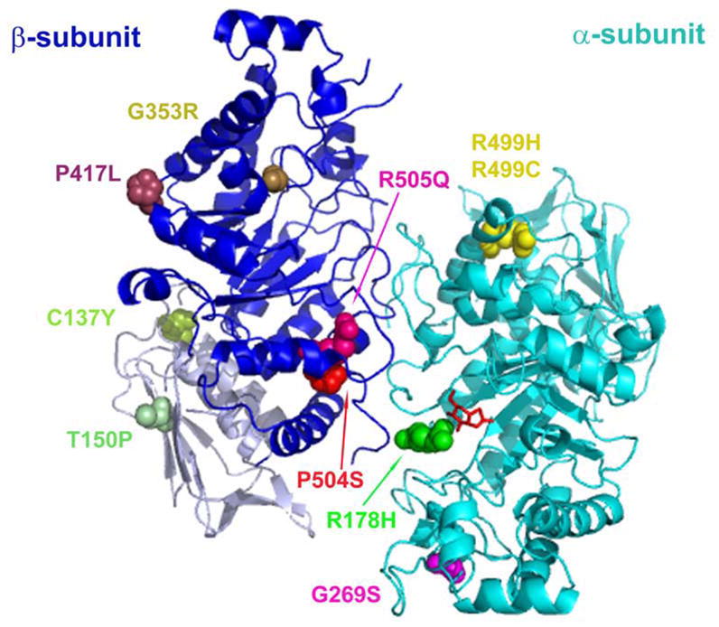
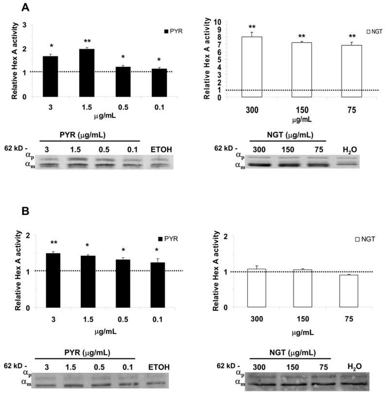
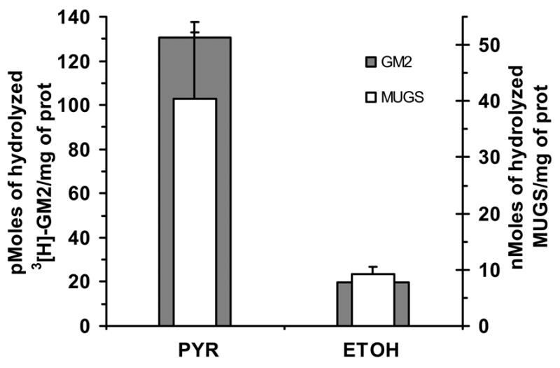
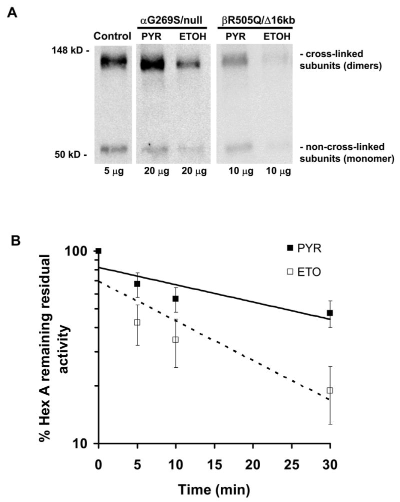
Late-onset GM2 gangliosidosis is composed of two related, autosomal recessive, neurodegenerative diseases, both resulting from deficiency of lysosomal, heterodimeric beta-hexosaminidase A (Hex A, alphabeta). Pharmacological chaperones (PC) are small molecules that can stabilize the conformation of a mutant protein, allowing it to pass the quality control system of the endoplasmic reticulum. To date all successful PCs have also been competitive inhibitors. Screening for Hex A inhibitors in a library of 1040 Food Drug Administration-approved compounds identified pyrimethamine (PYR (2,4-diamino 5-(4-chlorophenyl)-6-ethylpyrimidine)) as the most potent inhibitor. Cell lines from 10 late-onset Tay-Sachs (11 alpha-mutations, 2 novel) and 7 Sandhoff (9 beta-mutations, 4 novel) disease patients, were cultured with PYR at concentrations corresponding to therapeutic doses. Cells carrying the most common late-onset mutation, alphaG269S, showed significant increases in residual Hex A activity, as did all 7 of the beta-mutants tested. Cells responding to PC treatment included those carrying mutants resulting in reduced Hex heat stability and partial splice junction mutations of the inherently less stable alpha-subunit. PYR, which binds to the active site in domain II, was able to function as PC even to domain I beta-mutants. We concluded that PYR functions as a mutation-specific PC, variably enhancing residual lysosomal Hex A levels in late-onset GM2 gangliosidosis patient cells.


Figures

Fig. 1
Fig. 1
Replicate plot of a screen of the NINDS FDA-approved library of 1,040 small molecules for Hex A inhibitors. The graphic represents a replicate plot of residual Hex A activity in the presence of individual compounds from the NINDS library, i.e. each set of replicates define an X,Y coordinate. The zone outlined (dashed line) at 40% was set as the “inhibitor compound area,” which contained 4 compounds.
Fig. 2
Fig. 2
(A, B) Characterization of two inhibitors identified in the screen. The IC50 curves for the two “hits”, PYR (A) and thioguanine (TGN, B). (C) PYR behaves kinetically as a classical competitive inhibitor of Hex. (D) Maximal inhibition of Hex A by PYR was at pH 6.5, while the transition state analog, NGT, showed a maximum inhibition at pH of 4.5.
Fig. 3
Fig. 3
PYR enhances Hex A enzyme levels in some TSD patient cell lines. Two mutant TSD fibroblast cell lines, αG269S/IVS6+1G>A (A) and αR178H/R178H (B), were treated with PYR and NGT, at 20, 10 and 5 μg/mL, dissolved in DMSO. Hex A activities using MUGS as a substrate were determined. The relative fold-increase of Hex A activity was calculated based on the activity of cell lines treated only with DMSO. The mature (lysosomal) α-subunit protein levels seen in Western blots (beneath histograms) correlated with the relative increase of Hex A observed in the αG269S/IVS6+1G>A mutant (A). However, the cells containing the αR178H/R178H active site mutations failed to show any increases in relative Hex A activity or changes in α-subunit protein levels (B). αp, indicates the α-subunit precursor, αm, the mature lysosomal α-subunit. *P < 0.01, **P < 0.001.
Fig. 4
Fig. 4
Specificity of PYR as a PC for Hex A. Relative levels of MU-substrate hydrolysis by acid phosphatase (AcPhos, substrate MUP), Hex A (substrate, MUGS) and βGal (substrate, MUβGal) in one of TSD cell lines, αG269S/c1278insTACT, incubated with increasing concentrations of PYR (units mM).
Fig. 5
Fig. 5
Missense mutations identified in TSD and SD patients localized onto a three-dimensional Hex A ribbon diagram (2). Aminoacid residue alterations are highlighted in color and shown in a space filled format. Two novel mutations (βC137Y and βT150P) are located in domain I (βA50-P201; light gray) of the β-subunit (blue). NGT is shown as a red stick format bound in the active site of the α-subunit (cyan).
Fig. 6
Fig. 6
TSD or α-mutants cell lines which responded to PYR and NGT. The αG269S/c1278insTACT (A) and the αIVS9+1G>A/IVS8-7G>A (B) are shown with their relative Hex A activity and α-subunit protein levels at different treatment regimens of PYR and NGT. The fold-increase in activities was calculated based on the activity measured in control cell lines treated only with the dissolvent, i.e. ETOH for PYR-treated and H2O, for NGT-treated cells (in Methods). αp, indicates the α-subunit precursor, αm, the mature, lysosomal α-subunit. *P < 0.01, **P < 0.001.
Fig. 7
Fig. 7
Some TSD or α-mutants showed no relative increase in residual Hex A activity when exposed to PYR. PYR-treatment had no effect on cell lines containing αR499H, αR499C and αR178H mutations. The αR499H/c.1278insTACT mutant cell line (A) and αR178H/c.1510delC (B) are shown here in order to illustrate this observation. αp, indicates the α-subunit precursor, αm, the mature lysosomal α-subunit in Western blots.
Fig. 8
Fig. 8
PYR increases the enzyme activity and protein level of the α and β subunits of Hex A in the βR505Q/Δ16kb cell line. Histograms show relative activity enhancement of the lysate (total Hex A and S), as well as Hex A and S separated by IP (Methods). Western blots of the samples treated with PYR and NGT also document an increase in protein levels of the mature, lysosomal α- and β– subunits in the total lysate of treated cells. α/βp, indicates the α-and β-subunit precursors; αm and βm indicate the mature (lysosomal) α- and β-subunits, respectively. *P < 0.05; **P < 0.01.
Fig. 9
Fig. 9
Cellular localization of the mutant β-subunit-containing Hex isozymes in a patient fibroblast cell line, βR505Q/IVS11+5G>A. Cells were grown in the presence of PYR (3.0 μg/mL; A-F) or the solvent (ETOH) alone (control; G-L). The images in panels D-F represent selected areas of the same cell seen in panels A-C, but with higher magnification in order to demonstrate co-localization of the β-subunit of Hex (soluble protein in the lysosomal lumen) and Lamp-1 (integral lysosomal membrane protein) in lysosomes, versus the control cell, (G-I and J-L at higher magnification). Scale bars, red (10 μm) and magenta (2 μm).
Fig. 10
Fig. 10
Natural and artificial substrate activities are increased in a αG269S mutant TSD cell line grown in the presence of PYR (3 μg/mL). The level of Hex-enhancement as measured by natural substrate, 3[H]-GM2 ganglioside and recombinant Activator (gray bars), hydrolysis was similar to that observed with the α-active-site-specific artificial substrate, MUGS (white bars).
Fig. 11
Fig. 11
Effects of growth in PYR on the levels of monomeric and dimeric subunits of Hex; (A) Western blots from a normal, an α-G296S/null and a βR505Q/Δ16kb mutant cell line grown with or without PYR in their media. (B) Log scale of the percentage of remaining Hex A activity from βR505Q/Δ16kb mutant cell lysate exposed to PYR or solvent (ETOH) over a course of 30 minutes at 37°C.

Similar articles

Cited by

References

    1. Hirabayashi Y, Li YT, Li SC. J Neurochem. 1983;40(1):168–175. - PubMed
    1. Lemieux MJ, Mark BL, Cherney MM, Withers SG, Mahuran DJ, James MN. J Mol Biol. 2006;359(4):913–929. - PMC - PubMed
    1. Mark BL, Mahuran DJ, Cherney MM, Zhao D, Knapp S, James MN. J Mol Biol. 2003;327(5):1093–1109. - PMC - PubMed
    1. Sharma R, Bukovac S, Callahan J, Mahuran D. Biochim Biophys Acta. 2003;1637(1):113–118. - PMC - PubMed
    1. Hou Y, Tse R, Mahuran DJ. Biochemistry. 1996;35:3963–3969. - PubMed

Publication types