. 2007 Feb;9(2):149-59.
doi: 10.1038/ncb1527. Epub 2007 Jan 21.

Control of local actin assembly by membrane fusion-dependent compartment mixing

Affiliations

Control of local actin assembly by membrane fusion-dependent compartment mixing

Hoi-Ying E Yu et al. Nat Cell Biol. 2007 Feb.

Abstract

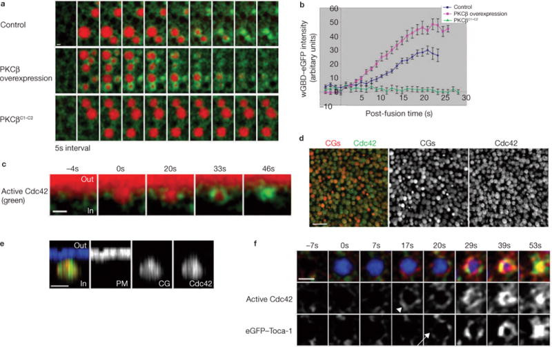
Local actin assembly is associated with sites of exocytosis in processes ranging from phagocytosis to compensatory endocytosis. Here, we examine whether the trigger for actin-coat assembly around exocytosing Xenopus egg cortical granules is 'compartment mixing'--the union of the contents of the plasma membrane with that of the secretory granule membrane. Consistent with this model, compartment mixing occurs on cortical granule-plasma membrane fusion and is required for actin assembly. Compartment mixing triggers actin assembly, at least in part, through diacylglycerol (DAG), which incorporates into the cortical granule membranes from the plasma membrane after cortical granule-plasma membrane fusion. DAG, in turn, directs long-term recruitment of protein kinase Cbeta (PKCbeta) to exocytosing cortical granules, where it is required for activation of Cdc42 localized on the cortical granules. The results demonstrate that mixing of two membrane compartments can direct local actin assembly and indicate that this process is harnessed during Xenopus egg cortical granule exocytosis to drive compensatory endocytosis.


Conflict of interest statement

COMPETING FINANCIAL INTERESTS

The authors declare that they have no competing financial interests.

Figures

Figure 1
Figure 1
Actin-coat assembly occurs after plasma membrane and cortical granule membrane incorporation on cortical granule exocytosis. (a) Subcortical view showing that actin coats (Alexa488–G-actin, arrowhead) assemble around the exocytosing cortical granules after cortical granule exocytosis (Texas Red–dextran, arrow). (b) z-view showing that actin (Utr1–261–mRFP) assembly occurs on cortical granule membranes (arrowhead) on cortical granule exocytosis (Alexa647–dextran) at 0 s, but not on the cortex (arrow). (c) Subcortical images showing that plasma membrane components (F-eGFP) are incorporated into the cortical granule membrane on cortical granule exocytosis (Alexa647–dextran, arrowhead), before actin-coat assembly (Utr1–261–mRFP, arrow). (d) z-view showing that plasma membrane (F-eGFP, green) incorporates into cortical granule membranes on cortical granule exocytosis (Texas Red–dextran) at 0 s. (e) Quantification of F-eGFP recruitment to exocytosing cortical granules (n = 16; 100% is maximum signal intensity before cortical granule constriction), where cortical granule exocytoses at 1.5 s. Signal intensity shown at 0 s represents basal signal intensity. The error bars represent s.e.m. (f, g) Subcortical images showing that BotA microinjection blocks cortical granule exocytosis (the absence of Texas Red–dextran filled compartment), and prevents (f) plasma membrane (F-eGFP) incorporation into the cortical granule membrane, as well as actin coat assembly (g; Alexa488–G-actin). InsP3 uncaging occurred at 0 s in a, f and g. Out, outside of cell; in, inside of cell. The scale bars represent 5 μm in a, f and g, 3 μm in b and 2 μm in c and d.
Figure 2
Figure 2
Ca2+ elevation triggers DAG generation on the plasma membrane, which incorporates into the cortical granule membrane on cortical granule–plasma membrane fusion. (a) z-view showing that DAG (eGFP–C1) is generated at the plasma membrane on Ca2+ increase and incorporates into the cortical granule membrane during cortical granule–plasma membrane fusion (Texas Red–dextran, arrowhead). (b) Subcortical view showing that DAG (eGFP–C1) is recruited to exocytosing cortical granules only (Texas Red–dextran, arrow), but not to non-exocytosed cortical granules (arrowhead). (c) Subcortical view showing that eGFP–C1mut (non-DAG binding) is not recruited to exocytosing cortical granules (arrowhead). InsP3 uncaging occurred at 0 s in a, b and c. The scale bars represent 2 μm in a and 4 μm in b and c.
Figure 3
Figure 3
DAG incorporation into cortical granule membranes occurs before actin assembly and is dependent on cortical granule–plasma membrane compartment mixing. (a) Subcortical view showing that DAG (mRFP–C1, red) incorporates into the cortical granule membrane when a cortical granules exocytoses at 0 s, before actin assembly (Alexa488–G-actin, green). (b) Quantification of eGFP–C1 recruitment to exocytosing cortical granules (n = 16; 100% is maximum signal intensity before cortical granule constriction), where the cortical granule exocytoses at 1.5 s. Signal intensity shown at 0 s represents basal signal intensity. The error bars represent s.e.m. (c) z-view showing that DAG (eGFP–C1) levels at the plasma membrane increases on InsP3 uncaging at 0 s, even though it fails to be incorporated into the cortical granule membrane when treated with BotA. The scale bars represent 1 μm in a and 2 μm in c.
Figure 4
Figure 4
DAG is required for actin-coat assembly. (a) z-view showing that C2 ceramide treatment inhibits DAG (eGFP–C1) elevation at the plasma membrane on Ca2+ increase. (b) Quantification of DAG intensity on plasma membrane showing the percentage increase in DAG levels after Ca2+ increase when compared with pre-activation levels (n = 12; asterisk indicates P = 0.00047). The error bars represent s.e.m. (c) Subcortical images showing that C2 ceramide treatment reduces actin coat (Alexa488–G-actin) assembly around exocytosing cortical granules (Texas Red–dextran), whereas DGK inhibitor enhances actin-coat assembly. (d) Quantification of the intensity of actin coats at exocytosing cortical granules (n = 20 for control; n = 21 for C2 ceramide; n = 21 for DGK inhibitor). The error bars represent s.e.m. (e) Exogenous addition of DAG induces cortical granule exocytosis (Alexa647–dextran) at 0 s, where PKCβ–eGFP is recruited to the exocytosing cortical granule before actin coat (Utr1–261–mRFP) assembly. The scale bars represent 1 μm in a and 2 μm in c and e.
Figure 5
Figure 5
PKCβ is transiently recruited to all cortical granules on Ca2+ elevation, but only remains on those that have exocytosed. (a) Schematic representation of the primary structure of PKCβ. The regulatory portion is comprised of C1 and C2 domains, which bind to DAG and phosphatidylserine and/or Ca2+, respectively. The catalytic portion is comprised of C3 and C4 domains, which bind to ATP and substrate, respectively. (b) Subcortical views showing that PKCβ–eGFP is recruited to all cortical granules on InsP3 uncaging at 0 s, but only remains on those that have fused with plasma membrane (Texas Red–dextran, arrow), and disappears from those that have not exocytosed (arrowhead). (c) z-view showing that PKCβ–eGFP disappears from cortical granule that has not exocytosed (the absence of a Texas Red–dextran filled compartment). (d) z-view showing that PKCβ–eGFP is translocated to the plasma membrane on InsP3 uncaging. (e) z-view showing that PKCβC1–C2–eGFP is present on the plasma membrane before InsP3 uncaging. (f) Subcortical views showing that PKCβC1–C2–eGFP is recruited to all cortical granules on InsP3 uncaging at 0 s, but only remains on those that have fused with plasma membrane (Texas Red–dextran, arrow), and disappears from those that have not exocytosed (arrowhead). (g) Subcortical images showing that eGFP–C2 is recruited to cortical granules (arrowhead) on InsP3 uncaging at 0 s before exocytosis. It subsequently disappears from cortical granules regardless of whether the cortical granules have exocytosed (Texas Red–dextran, arrow), or not (asterisk). (h) z-images showing that eGFP–C2 is translocated to plasma membrane on InsP3 uncaging. (i) Subcortical images showing that PKCβC1mut–eGFP (non-DAG binding PKCβ) is recruited to cortical granules (arrowhead) on InsP3 uncaging at 0 s before exocytosis. However, they disappear from cortical granules at later time points regardless of whether the cortical granules have exocytosed (Texas Red–dextran, arrow), or not (asterisk). (j) z-images showing that PKCβC1mut– eGFP is translocated to plasma membrane on InsP3 uncaging. The scale bars represent 4 μm in b, 2 μm in c, 5 μm in d, e and f, and 3 μm in gj.
Figure 6
Figure 6
Perturbation of PKCβ affects actin-coat assembly. (a) Subcortical images showing that overexpression of PKCβ induces formation of thicker, longer-lived actin coats (Alexa488–G-actin, arrows) on some exocytosing cortical granules (Texas Red–dextran), whereas others (arrowheads) have normal coats. In contrast, actin coats (Alexa488–G-actin) assemble normally (arrowheads) in PKCβK368R-overexpressing cells. InsP3 uncaging occurred at 0 s. (The background level of actin in the PKCβ-overexpressing figure is intentionally reduced so that the hyperdeveloped actin coats would not seem too saturated in green. Before adjustment, the actin background is comparable in both PKCβ-overexpressing and PKCβK368R-overexpressing cells.) (b) Subcortical images showing that exocytosing cortical granules (Texas Red–dextran) in cells expressing PKCβC1–C2 remain trapped at the plasma membrane over time with little or no actin coat (Alexa488–G-actin) assembly. Coexpression with full-length (FL) PKCβ partially rescues the phenotype, where assembly of actin coat (Alexa488–G-actin, arrowhead) is slightly delayed compare with control (arrowhead). (c) z-view showing the exocytosing cortical granule remains trapped at the plasma membrane when expressing PKCβC1–C2. (d) Quantification of time taken to retrieve cortical granule membranes (n = 117 for control; n = 90 for PKCβC1–C2; n = 89 for PKCβC1–C2with full-length PKCβ; P = 10−41 for control versus PKCβC1–C2; P = 10−25 for PKCβC1–C2 versus PKCβC1–C2 with full-length PKCβ). (e) Quantification of the intensity of actin coats at exocytosing cortical granules before coat compression. PKCβC1– C2 + FL1 and FL2 represent two levels of rescue in different cells. (n = 18 for control; n = 17 for PKCβC1–C2; n = 12 for PKCβC1–C2 with FL1; n = 5 for PKCβC1–C2 with FL2. The error bars represent s.e.m. in d and e. The scale bars represent 4 μm in a, 3 μm in b, and 2 μm in c.
Figure 7
Figure 7
Perturbation of PKCβ affects activation of Cdc42, which is localized on cortical granule membranes before egg activation. (a) Subcortical images showing that Cdc42 activation (wGBD–eGFP) on exocytosing cortical granules (Texas Red–dextran) is enhanced in cells overexpressing PKCβ, but reduced in cells expressing PKCβC1–C2. (b) Quantification of active Cdc42 recruitment to exocytosing cortical granules (n = 15 for control; n = 14 for PKCβ overexpression; n = 15 for PKCβC1–C2). The error bars represent s.e.m. (c) z-view showing that Cdc42 is activated (wGBD–eGFP) on cortical granule membrane when cortical granule exocytoses (Texas Red–dextran), but not on the cortex. (d) Low magnification of en face view showing immunofluorescence staining of Myc–Cdc42 (green) localizes on the cortical granules (red) before egg activation. (e) High magnification of z-view showing Myc–Cdc42 is localized only on cortical granules (red), but not on the plasma membrane (blue) before egg activation. (f) eGFP–Toca-1 (arrow) is recruited to the exocytosing cortical granule (Alexa647–dextran) after Cdc42 activation (wGBD–RFP, arrowhead). 0 s indicates time at which cortical granule exocytoses in c and f. The scale bars represent 1 μm in a, c and e, 10 μm in d and 2 μm in f.
Figure 8
Figure 8
Schematic representation of how compartment mixing results in actin-coat assembly. (a) Timeline showing the recruitment of different signalling molecules (black) with respect to the different events (red) during cortical granule (CG) exocytosis or endocytosis. (b) On calcium (green dot) increase, DAG (orange) is generated in the plasma membrane (PM, light-green line). Calcium binding results in the translocation of PKCβ (red) to the cortical granule (blue) and plasma membrane. On cortical granule–plasma membrane fusion, DAG from the plasma membrane incorporates into the cortical granule membranes, which sustains the recruitment of PKCβ at the cortical granule membrane. (c) Schematic representation summarizing how calcium elevation triggers different events that lead to actin-coat assembly (based on the results presented here and in ref. 4).

Similar articles

Cited by

References

    1. May RC, Machesky LM. Phagocytosis and the actin cytoskeleton. J Cell Sci. 2001;114:1061–1077. - PubMed
    1. Pollard TD, Borisy GG. Cellular motility driven by assembly and disassembly of actin filaments. Cell. 2003;112:453–465. - PubMed
    1. Bretscher MS, Aguado-Velasco C. Membrane traffic during cell locomotion. Curr Opin Cell Biol. 1998;10:537–541. - PubMed
    1. Sokac AM, Co C, Taunton J, Bement W. Cdc42-dependent actin polymerization during compensatory endocytosis in Xenopus eggs. Nature Cell Biol. 2003;5:727–732. - PubMed
    1. Nemoto T, Kojima T, Oshima A, Bito H, Kasai H. Stabilization of exocytosis by dynamic F-actin coating of zymogen granules in pancreatic acini. J Biol Chem. 2004;279:37544–37550. - PubMed

Publication types