Comparative Study
. 2007 Feb;13(2):204-10.
doi: 10.1038/nm1536. Epub 2007 Jan 21.

Angiotensin II type 1 receptor blockade attenuates TGF-beta-induced failure of muscle regeneration in multiple myopathic states

Affiliations
Comparative Study

Angiotensin II type 1 receptor blockade attenuates TGF-beta-induced failure of muscle regeneration in multiple myopathic states

Ronald D Cohn et al. Nat Med. 2007 Feb.

Erratum in

  • Nat Med. 2007 Apr;13(4):511

Abstract

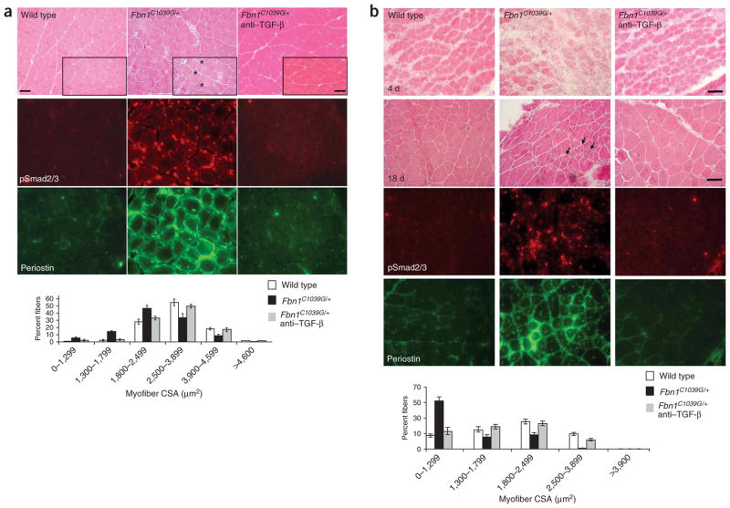
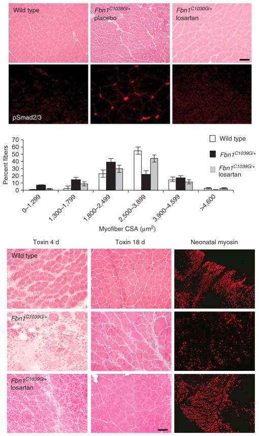
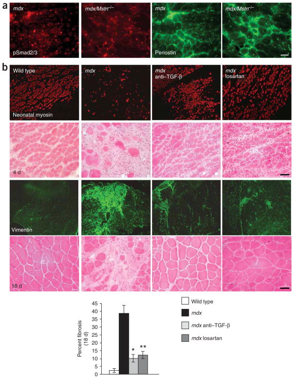
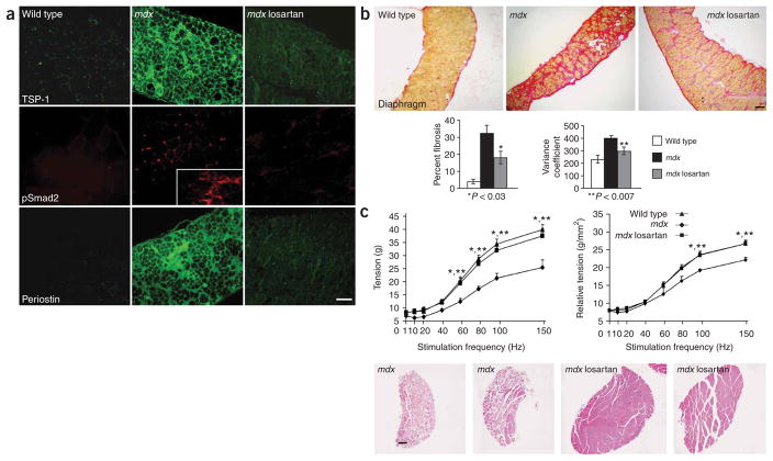
Skeletal muscle has the ability to achieve rapid repair in response to injury or disease. Many individuals with Marfan syndrome (MFS), caused by a deficiency of extracellular fibrillin-1, exhibit myopathy and often are unable to increase muscle mass despite physical exercise. Evidence suggests that selected manifestations of MFS reflect excessive signaling by transforming growth factor (TGF)-beta (refs. 2,3). TGF-beta is a known inhibitor of terminal differentiation of cultured myoblasts; however, the functional contribution of TGF-beta signaling to disease pathogenesis in various inherited myopathic states in vivo remains unknown. Here we show that increased TGF-beta activity leads to failed muscle regeneration in fibrillin-1-deficient mice. Systemic antagonism of TGF-beta through administration of TGF-beta-neutralizing antibody or the angiotensin II type 1 receptor blocker losartan normalizes muscle architecture, repair and function in vivo. Moreover, we show TGF-beta-induced failure of muscle regeneration and a similar therapeutic response in a dystrophin-deficient mouse model of Duchenne muscular dystrophy.


Conflict of interest statement

COMPETING INTERESTS STATEMENT

The authors declare that they have no competing financial interests.

Figures

Figure 1
Figure 1
Evaluation of steady-state and regenerating skeletal muscles in fibrillin-1 deficient mice. (a) Hematoxylin and eosin staining of quadriceps muscle (upper panels) shows marked variation of fiber size in Fbn1C1039G/+ mice. Note several small and split fibers (asterisks), fibers with central nucleation and endomysial thickening. TGF-β antagonism in vivo reverses myopathic architecture in Fbn1C1039G/+ mice. Increased TGF-β signaling, as evidenced by nuclear accumulation of pSmad2/3 (middle panels) and periostin expression (lower panels) in Fbn1C1039G/+ mice, when compared to wild-type mice or Fbn1C1039G/+ mice treated with TGF-β–neutralizing antibody. Analysis of the cross-sectional area (myofiber CSA in μm2) of tibialis anterior muscle fibers shows a decrease in fiber size in Fbn1C1039G/+ mice when compared to wild-type mice or Fbn1C1039G/+ mice treated with TGF-β–neutralizing antibody (graph). Scale bars, 70 μm (low magnification) and 50 μm (high magnification, inset boxes). (b) Impaired muscle regeneration in Fbn1C1039G/+ mice. Few newly-formed muscle fibers and disorganized muscle architecture with numerous small fibers (arrows) 4 and 18 days after cardiotoxin-induced muscle injury, respectively, in Fbn1C1039G/+ mice, when compared to wild-type mice or Fbn1C1039G/+ mice treated with TGF-β–neutralizing antibody (top panels). Increased TGF-β signaling, as evidenced by increased nuclear accumulation of pSmad2/3 and periostin expression in Fbn1C1039G/+ mice, when compared to wild-type mice or mice treated with TGF-β–neutralizing antibody (bottom panels). Morphometric analyses of tibialis anterior muscle 18 d after cardiotoxin injection shows reduced myofiber CSA (in μm2) in Fbn1C1039G/+ mice, when compared to wild-type mice or Fbn1C1039G/+ mice treated with TGF-β–neutralizing antibody (graph). Scale bars, 40 μm.
Figure 2
Figure 2
Losartan antagonizes TGFβ and restores muscle architecture and regeneration in Fbn1C1039G/+ mice. Placebo-treated Fbn1C1039G/+ mice show altered quadriceps steady-state muscle architecture with smaller fiber size and nuclear accumulation of pSmad2/3, when compared to wild-type or losartan-treated Fbn1C1039G/+ mice (top panels). Scale bar, 70 μm. Morphometric analyses showed reduced muscle fiber CSA (in μm2) in Fbn1C1039G/+ mice, when compared to wild-type or losartan-treated Fbn1C1039G/+ mice (graph). Few newly-formed muscle fibers with low neonatal myosin expression and disorganized muscle architecture 4 and 18 days after cardiotoxin-induced muscle injury, respectively, in Fbn1C1039G/+ mice, when compared to wild-type or losartan-treated Fbn1C1039G/+ mice (bottom panels). Scale bar, 40 μm.
Figure 3
Figure 3
Increased TGF-β signaling contributes to impaired muscle regeneration in mdx mice. (a) Increased nuclear accumulation of pSmad2/3 and sarcolemmal and extracellular matrix expression of periostin in dystrophin-deficient mdx mice (left panels) and mice deficient in both dystrophin and myostatin (mdx/Mstn−/−; right panels). Scale bar, 60 μm. (b) Reduced new fiber formation and neonatal myosin expression 4 days after cardiotoxin-induced injury in mdx mice, when compared to wild-type mice or mdx mice treated with TGF-β–neutralizing antibody or losartan (top panels). Extensive fibrosis (as evidenced by vimentin staining) and disorganized muscle architecture 18 days after muscle injury in mdx mice, when compared to wild-type mice or mdx mice treated with TGF-β–neutralizing antibody or losartan (bottom panels). The graph shows the percentage of fibrotic area as compared to the total area of muscle tissue 18 days after cardiotoxin injection. *P < 0.007 for mdx mice treated with TGF-β–neutralizing antibody versus untreated mdx mice; **P < 0.005 for losartan-treated versus untreated mdx mice. Scale bars, 40 μm.
Figure 4
Figure 4
Losartan decreases angiotensin II-mediated TGF-β signaling and improves muscle function in mdx mice. (a) Immunofluorescence analysis of target proteins downstream of the AT1. The diaphragm of mdx mice shows increased expression of thrombospondin-1 (TSP-1), a potent activator of TGF-β, and increased nuclear accumulation of pSmad2 and sarcolemmal and matrix expression of peripostin, when compared to wild-type or losartan-treated mdx mice. The inset box shows nuclear accumulation of pSmad2 in a connective tissue-rich region of diseased muscle. Scale bar, 100 μm. (b) Long-term losartan treatment attenuated myopathic disease progression in 9 month-old mdx mice. Representative sections of van Gieson–stained diaphragm from wild-type, mdx and losartan-treated mdx mice. Losartan-treated mice showed significantly (P 0.03) less fibrosis (red) than untreated mdx mice. Scale bar, 150 μm. The graphs show quantification of fibrotic area, expressed as percentage of total muscle area (left), and minimal Feret’s diameter variance coefficient (right). (c) In vitro force-frequency relationship of explanted EDL muscle. Isometric tension (g) versus stimulation frequency (1–150 Hz) was reduced in mdx mice at frequencies equal to or greater than 60 Hz, but was fully restored in losartan-treated mdx mice, as compared to wild-type animals (*wild-type versus mdx mice, **losartan-treated versus untreated mdx mice, P < 0.05; left graph). When force was normalized to muscle cross-sectional area (relative tension), losartan-treated mdx and wild-type mice were indistinguishable, whereas untreated mdx mice showed a significant decrease at frequencies of 100 and 150 Hz, when compared to either wild-type or losartan-treated mdx mice, (*wild-type versus mdx mice, **losartan-treated versus untreated mdx mice, P < 0.05; right graph). Representative low-power whole-muscle montages of EDL muscles from two untreated mdx mice (left) showed an overall decrease in muscle size and fiber content and an increase in fibrosis, as compared to two losartan-treated mdx mice (right). Wild-type mice, n = 4; untreated mdx mice, n = 3; losartan-treated mdx mice, n = 4. Scale bar, 150 μm.

Comment in

  • ACE inhibitor bulks up muscle.
    Chamberlain JS. Chamberlain JS. Nat Med. 2007 Feb;13(2):125-6. doi: 10.1038/nm0207-125. Nat Med. 2007. PMID: 17290265 No abstract available.

References

    1. Charge SB, Rudnicki MA. Cellular and molecular regulation of muscle regeneration. Physiol Rev. 2004;84:209–238. - PubMed
    1. Ng CM, et al. TGF-beta-dependent pathogenesis of mitral valve prolapse in a mouse model of Marfan syndrome. J Clin Invest. 2004;114:1586–1592. - PMC - PubMed
    1. Neptune ER, et al. Dysregulation of TGF-beta activation contributes to pathogenesis in Marfan syndrome. Nat Genet. 2003;33:407–411. - PubMed
    1. Allen RE, Boxhorn LK. Inhibition of skeletal muscle satellite cell differentiation by transforming growth factor-beta. J Cell Physiol. 1987;133:567–572. - PubMed
    1. Olson EN, Sternberg E, Hu JS, Spizz G, Wilcox C. Regulation of myogenic differentiation by type beta transforming growth factor. J Cell Biol. 1986;103:1799–1805. - PMC - PubMed

Publication types

MeSH terms