Comparative Study
. 2007 Feb;13(2):211-7.
doi: 10.1038/nm1488. Epub 2007 Jan 12.

Self-antigen tetramers discriminate between myelin autoantibodies to native or denatured protein

Affiliations
Comparative Study

Self-antigen tetramers discriminate between myelin autoantibodies to native or denatured protein

Kevin C O'Connor et al. Nat Med. 2007 Feb.

Abstract

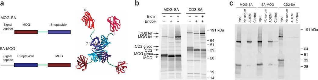
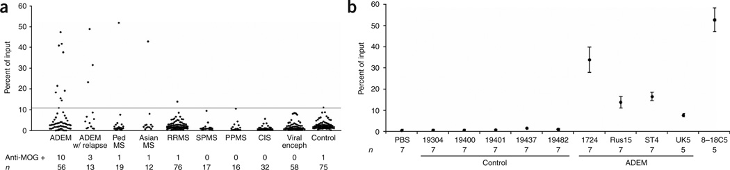
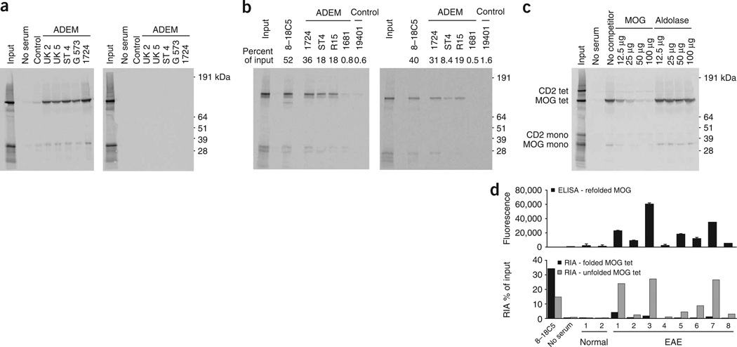
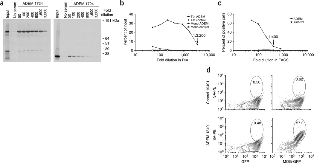
The role of autoantibodies in the pathogenesis of multiple sclerosis (MS) and other demyelinating diseases is controversial, in part because widely used western blotting and ELISA methods either do not permit the detection of conformation-sensitive antibodies or do not distinguish them from conformation-independent antibodies. We developed a sensitive assay based on self-assembling radiolabeled tetramers that allows discrimination of antibodies against folded or denatured myelin oligodendrocyte glycoprotein (MOG) by selective unfolding of the antigen domain. The tetramer radioimmunoassay (RIA) was more sensitive for MOG autoantibody detection than other methodologies, including monomer-based RIA, ELISA or fluorescent-activated cell sorting (FACS). Autoantibodies from individuals with acute disseminated encephalomyelitis (ADEM) selectively bound the folded MOG tetramer, whereas sera from mice with experimental autoimmune encephalomyelitis induced with MOG peptide immunoprecipitated only the unfolded tetramer. MOG-specific autoantibodies were identified in a subset of ADEM but only rarely in adult-onset MS cases, indicating that MOG is a more prominent target antigen in ADEM than MS.


Figures

Figure 1
Figure 1. Generation of tetrameric antigens for RIA
(a) Design of antigen-streptavidin fusion proteins. The extracellular Ig domain of MOG was connected to the N terminus (MOG-SA) or C terminus (SA-MOG) of the streptavidin monomer via a flexible linker. Crystal structures of MOG and streptavidin (PDB numbers PY9 and 1SW) were used to model the MOG-SA structure (right; MOG domains in red, streptavidin domains in blue). The N and C termini of the streptavidin monomer are solvent exposed on the same face, which enabled attachment of the antigen at either site. (b) Assembly of MOG-SA and CD2-SA fusion proteins into tetramers. 35S-labeled proteins were expressed in an in vitro translation system with endoplasmic reticulum microsomes to enable folding in a native environment. Both proteins were glycosylated, indicated by a decrease in molecular weight following digestion with EndoH (lanes 2 and 5). Addition of biotin during translation yielded tetramers stable during SDS-PAGE (lanes 3 and 6). (c) Specific immunoprecipitation of MOG tetramers by autoantibodies in serum from an ADEM individual (1724). Immunoprecipitation reactions were performed with MOG-SA, SA-MOG and CD2-SA tetramers using no serum, ADEM serum or a control serum at a dilution of 1:100. Data were standardized by calculating the percent of radiolabeled tetramer isolated in the immunoprecipitation as compared to the amount of input radiolabeled protein.
Figure 2
Figure 2. Analysis of MOG autoantibodies in CNS diseases
(a) The tetramer assay was used to examine serum samples from cases of ADEM (n = 56), ADEM with relapse (n = 13), different forms of MS (pediatric MS, n = 19; Asian MS, n = 12; relapsing-remitting MS, n = 76; secondary progressive MS, n = 17; primary progressive MS, n = 16) and clinically isolated syndrome (n = 32). Sera from cases of viral encephalitis (n = 58) and healthy individuals (n = 75) were used as controls. Each sample was assayed at a dilution of 1:100. Samples that precipitated >10.75% of the input MOG tetramer were considered positive. This threshold is 4 s.d. above the mean of healthy donors. No sample precipitated the CD2 control tetramer. (b) Results obtained in the MOG tetramer RIA were reproducible. Mean percentages and s.d. of immunoprecipitated MOG tetramer are indicated for 5–7 repeat measurements for sera from five healthy controls and four ADEM cases. Results with the 8–18C5 monoclonal antibody (positive control) are also shown.
Figure 3
Figure 3. The tetramer RIA permits discrimination of antibodies directed against conformation-dependent and conformation-independent epitopes
(a) ADEM autoantibodies against MOG are strictly conformation dependent. The tetrameric structure was maintained but the Ig domain of MOG was unfolded by cleavage of the MOG disulfide bond with DTT (heating at 70 °C for 10 min) (right). DTT was added to the same final concentration in immunoprecipitation reactions with untreated tetramer (left). (b) MOG binding by ADEM autoantibodies and the 8–18C5 monoclonal antibody did not require the N-linked glycan. The N-linked glycan was removed by mutation of MOG Asn 31 to Asp (left, wild-type; right, glycan-deficient). Immunoprecipitations were performed with 8–18C5 monoclonal antibody (50 ng) and ADEM and control sera. (c) Competition for autoantibody binding to MOG tetramers. Serum from an ADEM individual (1724) was incubated for 3 h with the indicated amounts of unlabeled recombinant MOG monomer or control protein (aldolase), before addition of radiolabeled MOG tetramer. (d) Antibodies from mice immunized with MOG (35–55) peptide bound to unfolded but not folded MOG. Sera from two control mice and eight mice that had been immunized with MOG (35–55) peptide in complete Freund’s adjuvant (CFA) were analyzed by ELISA using purified, refolded MOG extracellular domain expressed in E. coli (top) or by RIA using folded and unfolded MOG tetramer (bottom). The RIA showed that serum antibodies from MOG (35–55)-immunized mice bound to unfolded but not folded MOG. Seven of the eight immunized mice had clinical EAE with a score of 2 or 3; only one mouse did not develop EAE (#6).
Figure 4
Figure 4. Comparison of tetramer RIA to other autoantibody assays
(a) 35S-labeled MOG-SA tetramers (left) and MOG monomers (right) were compared in immunoprecipitation experiments using serial dilutions of serum from ADEM individual 1724. (b) Quantification of immunoprecipitation with tetramer or monomer as percentage of input radiolabeled protein. Tetrameric and monomeric versions had the same number of radiolabeled methionine residues per chain because the linker was retained in the monomeric form. (c) Antibodies in the serum of ADEM individual 1724 also bound to MOG on the surface of cells transfected with a MOG-GFP fusion protein. The ADEM serum was positive at a serum dilution of 1:400; no staining was observed at any dilution with the control serum (from individual 19437). (d) Example of FACS analysis. Jurkat cell lines transfected with MOG-GFP (right) and a control GFP vector (left) were stained with a control serum (from individual 19401) and an ADEM serum (from individual 1840) at a 1:50 serum dilution. Bound antibodies were detected with biotinylated antibodies to human IgG and streptavidin-PE. The IgG+ gates were centered on the GFP-bright population and set such that < 0.5% of live cells from the GFP vector control cell line were within the gate. For the ADEM sample, the intensity of antibody staining correlated with the level of MOG-GFP expression. Primary FACS data for all positive sera are shown in Supplementary Figure 2 online.

Similar articles

Cited by

References

    1. Linington C, Bradl M, Lassmann H, Brunner C, Vass K. Augmentation of demyelination in rat acute allergic encephalomyelitis by circulating mouse monoclonal antibodies directed against a myelin/oligodendrocyte glycoprotein. Am. J. Pathol. 1988;130:443–454. - PMC - PubMed
    1. Schluesener HJ, Sobel RA, Linington C, Weiner HL. A monoclonal antibody against a myelin oligodendrocyte glycoprotein induces relapses and demyelination in central nervous system autoimmune disease. J. Immunol. 1987;139:4016–4021. - PubMed
    1. Genain CP, et al. Antibody facilitation of multiple sclerosis-like lesions in a nonhuman primate. J. Clin. Invest. 1995;96:2966–2974. - PMC - PubMed
    1. Hjelmstrom P, Juedes AE, Fjell J, Ruddle NH. B-cell-deficient mice develop experimental allergic encephalomyelitis with demyelination after myelin oligodendrocyte glycoprotein sensitization. J. Immunol. 1998;161:4480–4483. - PubMed
    1. Berger T, et al. Antimyelin antibodies as a predictor of clinically definite multiple sclerosis after a first demyelinating event. N. Engl. J. Med. 2003;349:139–145. - PubMed

Publication types

MeSH terms