. 2006 Oct;2(10):e107.
doi: 10.1371/journal.ppat.0020107.

Quantitative membrane proteomics reveals new cellular targets of viral immune modulators

Affiliations

Quantitative membrane proteomics reveals new cellular targets of viral immune modulators

Eric Bartee et al. PLoS Pathog. 2006 Oct.

Abstract

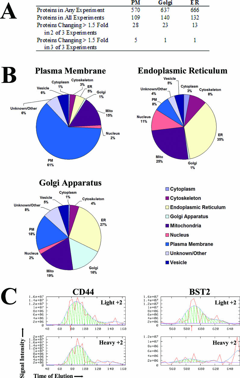
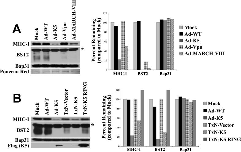
Immunomodulators of pathogens frequently affect multiple cellular targets, thus preventing recognition by different immune cells. For instance, the K5 modulator of immune recognition (MIR2) from Kaposi sarcoma-associated herpesvirus prevents activation of cytotoxic T cells, natural killer cells, and natural killer T cells by downregulating major histocompatibility complex (MHC) class I molecules, the MHC-like molecule CD1, the cell adhesion molecules ICAM-1 and PECAM, and the co-stimulatory molecule B7.2. K5 belongs to a family of viral- and cellular-membrane-spanning RING ubiquitin ligases. While a limited number of transmembrane proteins have been shown to be targeted for degradation by this family, it is unknown whether additional targets exist. We now describe a quantitative proteomics approach to identify novel targets of this protein family. Using stable isotope labeling by amino acids, we compared the proteome of plasma, Golgi, and endoplasmic reticulum membranes in the presence and absence of K5. Mass spectrometric protein identification revealed four proteins that were consistently underrepresented in the plasma membrane of K5 expression cells: MHC I (as expected), bone marrow stromal antigen 2 (BST-2, CD316), activated leukocyte cell adhesion molecule (ALCAM, CD166) and Syntaxin-4. Downregulation of each of these proteins was independently confirmed by immunoblotting with specific antibodies. We further demonstrate that ALCAM is a bona fide target of both K5 and the myxomavirus homolog M153R. Upon exiting the endoplasmic reticulum, ALCAM is ubiquitinated in the presence of wild-type, but not RING-deficient or acidic motif-deficient, K5, and is targeted for lysosomal degradation via the multivesicular body pathway. Since ALCAM is the ligand for CD6, a member of the immunological synapse of T cells, its removal by viral immune modulators implies a role for CD6 in the recognition of pathogens by T cells. The unbiased global proteome analysis therefore revealed novel immunomodulatory functions of pathogen proteins.


Conflict of interest statement

Competing interests. The authors have declared that no competing interests exist.

Figures

Figure 1
Figure 1. Schematic Representation of the SILAC Labeling and Purification Protocol
HeLa cells were grown for 5 d in labeling medium to ensure complete labeling. They were then infected with either adenovirus vector (light sample) or adenovirus-expressing K5 (heavy sample). Then 24 h post-infection, cells were harvested and counted, and equal numbers of cells combined. Samples were lysed in a Dounce homogenizer and unlysed cells removed by centrifugation. Membrane and soluble proteins were separated by centrifugation. The membrane pellet was resuspended, and different membrane fractions were separated over a discontinuous sucrose gradient. The bands corresponding to the plasma membrane (PM), Golgi, and ER fractions were removed and the proteins pelleted. The resulting pellet was then washed with sodium carbonate to remove non-integral membrane proteins, followed by ammonium bicarbonate to remove salts and other impurities. The final pellet was resuspended in 8 M urea and digested with trypsin for MS/MS analysis.
Figure 2
Figure 2. Protein Identification and Differential Peptide Quantification
(A) Approximately 500–700 proteins were identified by at least one peptide in each fraction; however, fewer than 150 proteins per fraction were identified in all three replicates. Of these, only five proteins changed more than 1.5-fold in the plasma membrane (PM) fraction, while only one protein changed more than 1.5-fold in all three replicates of either the Golgi or ER fractions. (B) Proteins present in all three replicates of each fraction were analyzed for their predicted subcellular distribution using annotations in the Swiss-Prot proteomics database. (C) Comparison of signal intensities obtained for selected peptides from the indicated proteins. The red line indicates the actual raw data recovered from the mass spectrometer; the blue line is a hypothetical best fit line drawn to help analyze elution time of the light and heavy peptides. The orange arrows indicate the time at which the initial MS/MS scan was initiated. Peptides derived from unaffected proteins, such as CD44, display a similar intensity for both the light and heavy isotopes. Peptides derived from differentially expressed proteins, such as BST-2, show clearly different intensity peaks for the light and heavy peptides. Note the slightly different scale for intensity of each peptide.
Figure 3
Figure 3. BST-2 Is Differentially Expressed in K5-Expressing Cells
HeLa cells were transduced with Ad-WT, Ad-K5, Ad-vpu, or Ad-MARCH-VIII. Then 24 h post-infection, cells were harvested and whole cell lysates were analyzed for the abundance of BST-2 and MHC I by Western blotting. Note that BST-2 is highly glycosylated and runs as multiple bands. The high molecular weight band marked with an asterisk is non-specific. Equal protein loading was confirmed by visualizing the ER resident chaperone Bap31 as well as general protein staining with Ponceau red ([A], left). The intensity of each band was quantified using densitometry ([A], right). The specificity of BST-2 downregulation was confirmed by transfecting K5 or a catalytically inactive K5-RING mutant, which showed no effect on either MHC I or BST-2 (B). The protein bands corresponding to BST-2 are more intense in (B) than in (A) because of longer exposure of the autoradiograph as well as variation in electrophoretic separation. Note that the same lysates were analyzed in lanes 1–3 of the blots in both (A) and (B).
Figure 4
Figure 4. K5 Causes Downregulation but Not Relocalization of Syntaxin-4
(A) HeLa cells were transduced with Ad-WT, Ad-K5, Ad-vpu, or Ad-MARCH-VIII. Then 24 h post-infection, cells were harvested and whole cell lysates were analyzed as in Figure 3, except that antibodies specific for Syntaxin-4 were used. Expression of K5 resulted in a moderate but reproducible reduction of Syntaxin-4 levels. (B) To determine if K5 or MARCH proteins caused a relocalization of Syntaxin-4, HeLa cells were transfected with C-terminally FLAG-tagged versions of the E3 enzymes shown. Cells were then analyzed for the location of Syntaxin-4 (using an Alexa Fluor 594–conjugated secondary antibody, shown as red) as well as the overexpressed E3 (using a FITC-conjugated anti-FLAG antibody, shown as green). Co-localization of Syntaxin-4 and the ubiquitin ligases is revealed as yellow co-staining. While K5 did not co-localize with Syntaxin-4, several MARCH family proteins relocalized Syntaxin-4 to a MARCH-containing compartment. A naturally occurring splice variant of MARCH-IX that lacks a complete RING domain and fails to exit the ER was unable to relocalize Syntaxin-4. (C) C-terminal truncation mutants of MARCH-VIII that failed to exit the ER do not co-localize with Syntaxin-4.
Figure 5
Figure 5. K5 Downregulates ALCAM
(A) C-terminally HA-tagged ALCAM was co-transfected with K5 or control plasmid. Then 24 h post-transfection, cells were harvested and the abundance of ALCAM-HA in each lysate was measured by Western blotting with anti-HA antibody. (B) Cell surface expression of endogenous ALCAM in the presence or absence of K5 was determined by flow cytometry. HeLa cells were co-transfected with K5 and a green fluorescent protein (GFP)–expressing plasmid to identify transfectants. Then 24 h post-transfection, cells were harvested and stained with antibodies against MHC I, ALCAM, CD9, and CD29. Both vector (solid black) and K5-transfected (white) cells were gated for GFP-expressing cells. (C) ALCAM downregulation by K5-related proteins was determined by transfecting HeLa cells with viral K3 family members (K3 and M153R) and the indicated human MARCH proteins. Also examined were mutant K5 proteins with enzymatically inactive RING-CH domains (K5-RING) or lacking acidic residues implicated in subcellular targeting (K5DE12). Neither K5 mutant reduced ALCAM levels. KSHV K3 was unable to downregulate ALCAM, whereas the myxomavirus M153R protein significantly reduced ALCAM surface expression. Two of the MARCH proteins, MARCH-IV and MARCH-IX, strongly downregulated ALCAM, while MARCH-VIII showed a minimal effect. (D) To determine whether ALCAM expression was affected by KSHV, latently infected immortalized DMVECs were treated with PMA to induce expression of lytic genes including K5. Surface levels of either MHC I or ALCAM were measured by flow cytometry 24 and 48 h post-induction. CD81, measured at 24 h, was used as a control. The ratio between the mean fluorescence intensity of infected and uninfected samples from these experiments is shown as fold change on the right.
Figure 6
Figure 6. Ubiquitination and Lysosomal Degradation of ALCAM in the Presence of K5
(A) Ubiquitination of ALCAM was examined by co-transfection of HeLa cells with ALCAM-HA as well as the indicated E3 enzymes. Then 24 h post-transfection, cells were lysed in 1% CHAPS, and ALCAM-HA was immunoprecipitated using anti-HA antibody. Samples were resolved on an 8% SDS-PAGE gel, transferred to PVDF, and immunoblotted (WB) with the anti-ubiquitin (Ubi) antibody P4D1 (top) or anti-HA (bottom). Ubiquitinated ALCAM was visible upon co-transfection of K5 and MARCH-VIII, but not with the inactive K5DE12 mutant and the unrelated HIV immune modulator vpu. (B) To determine whether ubiquitinated ALCAM was degraded by proteasomes or in lysosomes, the fate of newly synthesized ALCAM-HA was determined by metabolic labeling for 10 min with S35 Met/Cys and chasing the label for the indicated times (hours) in the presence of the indicated inhibitors. Following lysis, ALCAM-HA was immunoprecipitated using the HA antibody and samples were treated overnight with endoglycosidase H followed by electrophoretic separation. Note the increased recovery of ALCAM at 8 h in the presence of the endosomal/lysosomal proton pump inhibitor concanamycin A (ConA), but not in the presence of the proteasomal inhibitor MG132 (50 μmol). (C) Surface expression of ALCAM can be restored by overexpressing a dominant negative version of the AAA-ATPase Vps4, which is essential for targeting proteins to MVBs. HeLa cells were transfected as indicated together with GFP to identify transfected cells. Then 24 h post-transfection, cells were harvested and the surface expression of either MHC I or ALCAM was analyzed using flow cytometry. The graph shows the ratio of mean fluorescence intensity from K5-transfected cells to that of control cells after gating for GFP. Data are averaged from three separate experiments.

Similar articles

Cited by

References

    1. Wiertz EJ, Jones TR, Sun L, Bogyo M, Geuze HJ, et al. The human cytomegalovirus US11 gene product dislocates MHC class I heavy chains from the endoplasmic reticulum to the cytosol. Cell. 1996;84:769–779. - PubMed
    1. Johnson DC, Hegde NR. Inhibition of the MHC class II antigen presentation pathway by human cytomegalovirus. Curr Top Microbiol Immunol. 2002;269:101–115. - PubMed
    1. Ziegler H, Muranyi W, Burgert HG, Kremmer E, Koszinowski UH. The luminal part of the murine cytomegalovirus glycoprotein gp40 catalyzes the retention of MHC class I molecules. EMBO J. 2000;19:870–881. - PMC - PubMed
    1. Lodoen M, Ogasawara K, Hamerman JA, Arase H, Houchins JP, et al. NKG2D-mediated natural killer cell protection against cytomegalovirus is impaired by viral gp40 modulation of retinoic acid early inducible 1 gene molecules. J Exp Med. 2003;197:1245–1253. - PMC - PubMed
    1. Kerkau T, Bacik I, Bennink JR, Yewdell JW, Hunig T, et al. The human immunodeficiency virus type 1 (HIV-1) Vpu protein interferes with an early step in the biosynthesis of major histocompatibility complex (MHC) class I molecules. J Exp Med. 1997;185:1295–1305. - PMC - PubMed

Publication types

MeSH terms