. 2007 Jan 19;3(1):e11.
doi: 10.1371/journal.pcbi.0030011. Epub 2006 Dec 8.

Evolutionary interactions between N-linked glycosylation sites in the HIV-1 envelope

Affiliations

Evolutionary interactions between N-linked glycosylation sites in the HIV-1 envelope

Art F Y Poon et al. PLoS Comput Biol. .

Abstract

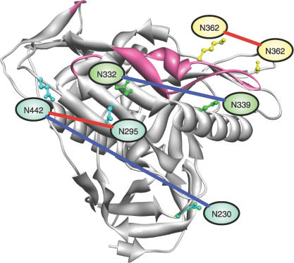
The addition of asparagine (N)-linked polysaccharide chains (i.e., glycans) to the gp120 and gp41 glycoproteins of human immunodeficiency virus type 1 (HIV-1) envelope is not only required for correct protein folding, but also may provide protection against neutralizing antibodies as a "glycan shield." As a result, strong host-specific selection is frequently associated with codon positions where nonsynonymous substitutions can create or disrupt potential N-linked glycosylation sites (PNGSs). Moreover, empirical data suggest that the individual contribution of PNGSs to the neutralization sensitivity or infectivity of HIV-1 may be critically dependent on the presence or absence of other PNGSs in the envelope sequence. Here we evaluate how glycan-glycan interactions have shaped the evolution of HIV-1 envelope sequences by analyzing the distribution of PNGSs in a large-sequence alignment. Using a "covarion"-type phylogenetic model, we find that the rates at which individual PNGSs are gained or lost vary significantly over time, suggesting that the selective advantage of having a PNGS may depend on the presence or absence of other PNGSs in the sequence. Consequently, we identify specific interactions between PNGSs in the alignment using a new paired-character phylogenetic model of evolution, and a Bayesian graphical model. Despite the fundamental differences between these two methods, several interactions are jointly identified by both. Mapping these interactions onto a structural model of HIV-1 gp120 reveals that negative (exclusive) interactions occur significantly more often between colocalized glycans, while positive (inclusive) interactions are restricted to more distant glycans. Our results imply that the adaptive repertoire of alternative configurations in the HIV-1 glycan shield is limited by functional interactions between the N-linked glycans. This represents a potential vulnerability of rapidly evolving HIV-1 populations that may provide useful glycan-based targets for neutralizing antibodies.


Conflict of interest statement

Competing interests. The authors have declared that no competing interests exist.

Figures

Figure 1
Figure 1. The Nature of Glycan Interactions Depends on Their Intramolecular Distance
Potential PNGSs that were identified by the disequon model as having significant interactions with other PNGSs were mapped to a structural model of HIV-1 gp120. Intramolecular distances between interacting PNGSs were measured using the visualization software UCSF Chimera [53]. When an asparagine residue corresponding to a PNGS was not present in the structural model, the distance between pairs was approximated by 4.0 Å times the number of residues separating the PNGSs, to a maximum of five residues. If the number of residues exceeded five, then the unmapped pair was omitted from the analysis.
Figure 2
Figure 2. Consensus Bayesian Network of Highly Polymorphic PNGSs in the HIV-1 Envelope
(A) Ordered distribution of frequencies that undirected links (i.e., arcs) occurred in replicate networks optimized on 100 random samples of 200 sequences from the alignment, expressed as a percentage (y-axis). A dotted line at 50% indicates an arbitrary cutoff, above which arcs were applied to assembling the consensus network. (B) Graph of consensus network. Each node (oval) represents the PNGS identified by its position in the HxB2 reference sequence, with one exception (V2ins) that represents an inserted PNGS downstream of the conserved N186. Arcs between nodes are labeled with percentage of occurrence above and odds-ratios below. An odds-ratio <1 (red) indicates mutual exclusion and >1 (blue) co-occurrence of PNGSs.
Figure 3
Figure 3. Interacting PNGS Mapped to Structural Model of gp120
Arcs from the consensus network are mapped to asparagine residues in the structure of a folded CD4-bound gp120 protein. The host cell-binding face of gp120 is oriented towards the top of the figure, and variable loops (of which many residues are truncated) are highlighted in pink. Arcs are colored red to indicate mutual exclusion, and blue indicates co-occurrence of the PNGSs. The asparagine residues of mutually exclusive PNGSs N295/N442 and N362/N465 are located within ∼12 Å of one another. In contrast, co-occurent PNGSs are separated by much greater intramolecular distances.
Figure 4
Figure 4. Disequon Model Parameters
This diagram illustrates the eight rates that are parameterized by the disequon (paired-character) model. Each box corresponds to a state in the disequon model, in which the first digit represents the presence or absence of a PNGS at the first site, and the second digit for the second site. The interaction parameter (ɛ) between paired sites is highlighted in red.

References

    1. Weerapana E, Imperiali B. Asparagine-linked protein glycosylation: From eukaryotic to prokaryotic systems. Glycobiology. 2006;16:91R–101R. - PubMed
    1. Gavel Y, von Heijne G. Sequence differences between glycosylated and non-glycosylated Asn-X-Thr/Ser acceptor sites: Implications for protein engineering. Protein Eng. 1990;3:433–442. - PMC - PubMed
    1. Chen W, Helenius J, Braakman I, Helenius A. Cotranslational folding and calnexin binding during glycoprotein synthesis. Proc Natl Acad Sci U S A. 1995;92:6229–6233. - PMC - PubMed
    1. Meunier JC, Fournillier A, Choukhi A, Cahour A, Cocquerel L, et al. Analysis of the glycosylation sites of the hepatitis C virus (HCV) glycoprotein E1 and the influence of E1 glycans on the formation of the HCV glycoprotein complex. J Gen Virol. 1999;80:887–896. - PubMed
    1. Goffard A, Callens N, Bartosch B, Wychowski C, Cosset FL, et al. Role of N-linked glycans in the functions of hepatitis C virus envelope glycoproteins. J Virol. 2005;79:8400–8409. - PMC - PubMed

Publication types

Associated data