Clinical Trial
. 2007 Jan;3(1):e4.
doi: 10.1371/journal.ppat.0030004.

In vitro derived dendritic cells trans-infect CD4 T cells primarily with surface-bound HIV-1 virions

Affiliations
Clinical Trial

In vitro derived dendritic cells trans-infect CD4 T cells primarily with surface-bound HIV-1 virions

Marielle Cavrois et al. PLoS Pathog. 2007 Jan.

Abstract

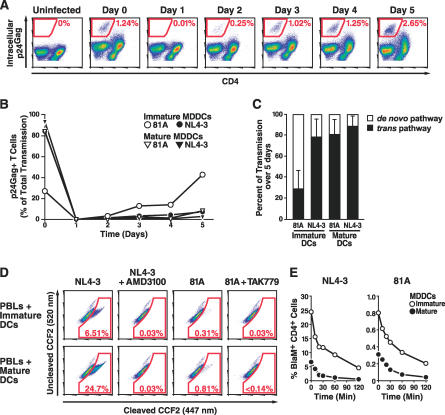
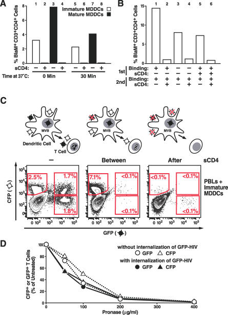
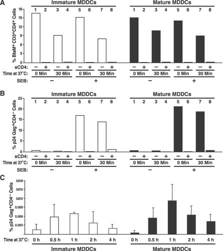
In the prevailing model of HIV-1 trans-infection, dendritic cells (DCs) capture and internalize intact virions and transfer these virions to interacting T cells at the virological synapse. Here, we show that HIV-1 virions transmitted in trans from in vitro derived DCs to T cells principally originate from the surface of DCs. Selective neutralization of surface-bound virions abrogated trans-infection by monocyte-derived DCs and CD34-derived Langerhans cells. Under conditions mimicking antigen recognition by the interacting T cells, most transferred virions still derived from the cell surface, although a few were transferred from an internal compartment. Our findings suggest that attachment inhibitors could neutralize trans-infection of T cells by DCs in vivo.


Conflict of interest statement

Competing interests. The authors have declared that no competing interests exist.

Figures

Figure 1
Figure 1. Mature MDDCs Transmit HIV-1 to T Cells Primarily via the trans Pathway
(A–C) NL4–3 or 81A virions were bound to MDDCs. After washing, the cells were added to activated autologous T cells immediately or after 1 to 5 d of culture at 37 °C to allow HIV-1 transmission to T cells for 24 h. The number of transmission events was measured by monitoring the appearance of infected T cells detected by p24Gag intracellular immunostaining. (A) Transmission of 81A virions from immature MDDCs to T cells over time. FACS plots represent the population of T cells (CD3+CD1a) analyzed for intracellular Gag and CD4 expression. (B) Effects of maturation on HIV-1 transmission from MDDCs to T cells. Values are percentages of all transmission events over 5 d. (C) Relative contribution of trans pathway or de novo pathway in transmission of virus from immature or mature MDDCs to T cells. Data are averaged from MDDCs derived from nine donors. Transmission events from days 1 to 5 were added to determine the number of transmission events occurring via the de novo pathway. (D and E) NL4–3 or 81A virions containing BlaM-Vpr were bound to MDDCs at 4 °C. After washing, the cells were added to autologous T cells immediately (T0) or after incubation for 10 to 120 min at 37 °C. HIV-1 transmission was measured with a virion-based fusion assay after gating on CD3+CD4+ cells. (D) HIV-1 transmission from MDDCs to T cells at T0. FACS plots show CD3+CD4+CD1a cells analyzed for virion fusion. To control for specificity, MDDCs were incubated with T cells and entry inhibitors (500 nM TAK-779 or 500 nM AMD3100). (E) Effect of time on NL4–3 and 81A transmission from MDDCs to T cells. The curve is representative of four experiments.
Figure 2
Figure 2. HIV-1 Virions Transmitted In Trans from MDDCs to T Cells Are Sensitive to sCD4 and Pronase
(A) Immature and mature MDDCs were incubated at 4 °C with NL4–3 virions containing BlaM-Vpr, washed, and incubated for 30 min at 37 °C to allow virion internalization or held at 4 °C. MDDCs were treated (or not) with sCD4 at 4 °C to inactivate cell-surface virions. After extensive washes, loaded MDDCs were added to autologous PBLs to allow HIV-1 trans-infection of T cells. Fusion was measured with the virion-based fusion assay after gating on the CD3+CD4+ cells. The histogram presents a representative experiment independently performed five times with similar results. (B) sCD4 treatment of MDDCs loaded with HIV-1 virions does not inhibit transfer of virions loaded after treatment. Two sets of HIV-1 virions containing BlaM-Vpr were successively bound to immature MDDCs and neutralized with sCD4. After incubation with autologous CD4 T cells, fusion to CD3+CD4+ cells was assessed. (C) MDDCs were sequentially incubated with two reporter viruses (1 μg of p24Gag each). GFP-HIV was bound and internalized, but CFP-HIV was bound only. Loaded MDDCs were incubated with autologous PBLs for 2 d. FACS plots show T cells analyzed for GFP and CFP expression when MDDCs were untreated or treated with sCD4 at 4 °C between or after loading of the reporter viruses. (D) GFP-HIV and CFP-HIV were either successively bound to MDDCs or GFP-HIV was, in addition, internalized by MDDCs during a 30-min incubation at 37 °C. MDDCs were then treated with increasing concentrations of pronase. Curves represent the number of T cells expressing CFP or GFP expressed as a percentage of the untreated samples. This graph presents a representative experiment with mature MDDCs that was independently performed three times with both immature and mature MDDCs.
Figure 3
Figure 3. HIV-1 Transmitted from SEB-Stimulated MDDCs to T Cells Remains Mostly Sensitive to sCD4
NL4–3 virions containing BlaM-Vpr were bound to SEB-pulsed MDDCs. The MDDCs were then incubated or not at 37 °C for the indicated time, treated or not with sCD4 at 4 °C, and incubated with purified autologous CD4 T cells. (A–C) Viral transfer was measured by analyzing HIV-1 fusion to CD4+CD3+ T cells after 2 h of culture (A) and by measuring productive infection of the T cells by intracellular Gag immunostaining 3 d later (B and C). (B) Histogram depicts one representative experiment performed three times with cells from three independent donors. (C) HIV-1 transfer to resting T cells from DCs pulsed with SEB and treated with sCD4 over time. This experiment was performed in triplicate; similar results obtained with MDDCs from two additional donors.

References

    1. Banchereau J, Steinman RM. Dendritic cells and the control of immunity. Nature. 1998;392:245–252. - PubMed
    1. Spira AI, Marx PA, Patterson BK, Mahoney J, Koup RA, et al. Cellular targets of infection and route of viral dissemination after an intravaginal inoculation of simian immunodeficiency virus into rhesus macaques. J Exp Med. 1996;183:215–225. - PMC - PubMed
    1. Hu J, Gardner MB, Miller CJ. Simian immunodeficiency virus rapidly penetrates the cervicovaginal mucosa after intravaginal inoculation and infects intraepithelial dendritic cells. J Virol. 2000;74:6087–6095. - PMC - PubMed
    1. Hu Q, Frank I, Williams V, Santos JJ, Watts P, et al. Blockade of attachment and fusion receptors inhibits HIV-1 infection of human cervical tissue. J Exp Med. 2004;199:1065–1075. - PMC - PubMed
    1. Cameron PU, Freudenthal PS, Barker JM, Gezelter S, Inaba K, et al. Dendritic cells exposed to human immunodeficiency virus type-1 transmit a vigorous cytopathic infection to CD4+ T cells. Science. 1992;257:383–387. - PubMed

Publication types