

Projection map of aquaporin-9 at 7 A resolution
- PMID: 17239399
- PMCID: PMC1839870
- DOI: 10.1016/j.jmb.2006.12.042
Projection map of aquaporin-9 at 7 A resolution
Abstract
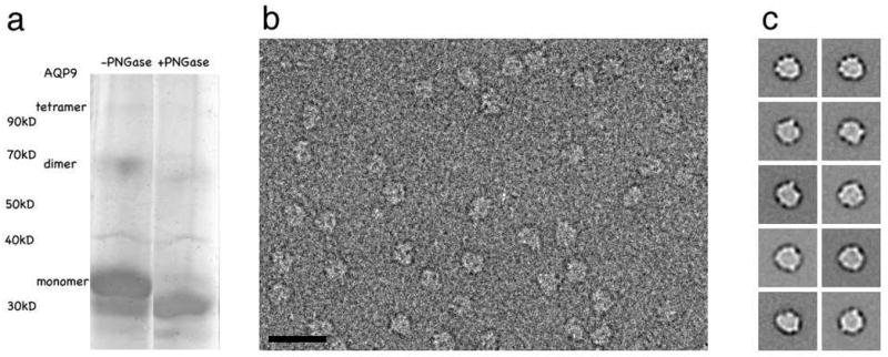
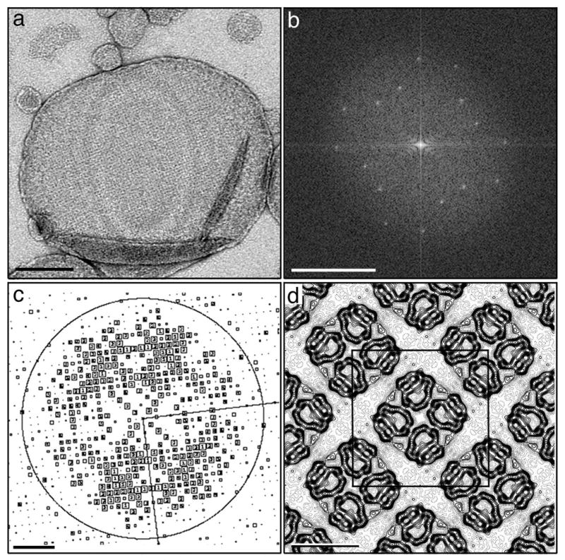
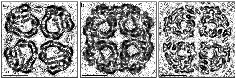
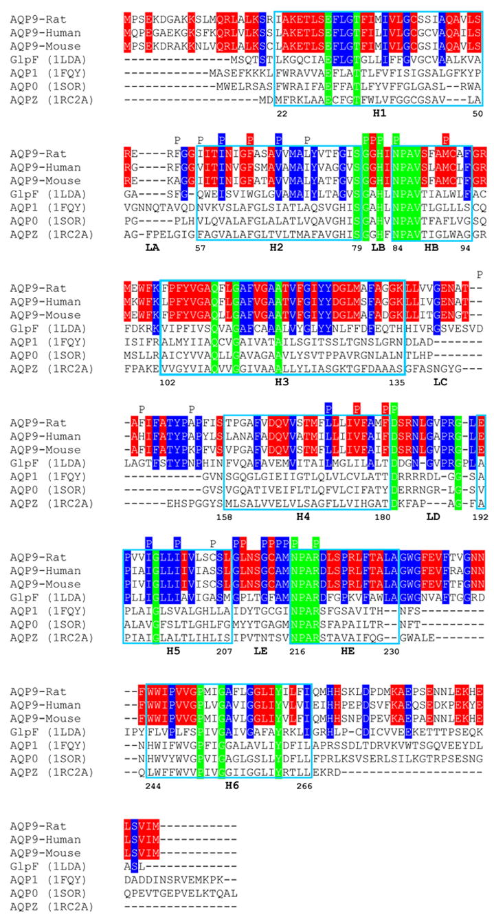
Aquaporin-9, an aquaglyceroporin present in diverse tissues, is unique among aquaporins because it is not only permeable to water, urea and glycerol, but also allows passage of larger uncharged solutes. Single particle analysis of negatively stained recombinant rat aquaporin-9 revealed a particle size characteristic of the tetrameric organization of all members of the aquaporin family. Reconstitution of aquaporin-9 into two-dimensional crystals enabled us to calculate a projection map at 7 A resolution. The projection structure indicates a tetrameric structure, similar to GlpF, with each square-like monomer forming a pore. A comparison of the pore-lining residues between the crystal structure of GlpF and a homology model of aquaporin-9 locates substitutions in these residues predominantly to the hydrophobic edge of the tripathic pore of GlpF, providing first insights into the structural basis for the broader substrate specificity of aquaporin-9.
Figures


Similar articles
-
The 6.9-A structure of GlpF: a basis for homology modeling of the glycerol channel from Escherichia coli.J Struct Biol. 2000 Nov;132(2):133-41. doi: 10.1006/jsbi.2000.4317. J Struct Biol. 2000. PMID: 11162735
-
The 3.7 A projection map of the glycerol facilitator GlpF: a variant of the aquaporin tetramer.EMBO Rep. 2000 Aug;1(2):183-9. doi: 10.1093/embo-reports/kvd022. EMBO Rep. 2000. PMID: 11265760 Free PMC article.
-
Water and glycerol permeation through the glycerol channel GlpF and the aquaporin family.J Synchrotron Radiat. 2004 Jan 1;11(Pt 1):86-8. doi: 10.1107/s0909049503023872. Epub 2003 Nov 28. J Synchrotron Radiat. 2004. PMID: 14646142 Review.
-
Architecture and selectivity in aquaporins: 2.5 a X-ray structure of aquaporin Z.PLoS Biol. 2003 Dec;1(3):E72. doi: 10.1371/journal.pbio.0000072. Epub 2003 Dec 22. PLoS Biol. 2003. PMID: 14691544 Free PMC article.
-
The structure of GlpF, a glycerol conducting channel.Novartis Found Symp. 2002;245:51-61; discussion 61-5, 165-8. Novartis Found Symp. 2002. PMID: 12027015 Review.
Cited by
-
Sample Preparation and Data Collection for Electron Crystallographic Studies on Membrane Protein Structures and Lipid-Protein Interaction.Methods Mol Biol. 2021;2215:247-265. doi: 10.1007/978-1-0716-0966-8_11. Methods Mol Biol. 2021. PMID: 33368007
-
Overview of electron crystallography of membrane proteins: crystallization and screening strategies using negative stain electron microscopy.Curr Protoc Protein Sci. 2013;Chapter 17:Unit17.15. doi: 10.1002/0471140864.ps1715s72. Curr Protoc Protein Sci. 2013. PMID: 23546618 Free PMC article.
-
Biogenesis of bacterial inner-membrane proteins.Cell Mol Life Sci. 2010 Jul;67(14):2343-62. doi: 10.1007/s00018-010-0303-0. Epub 2010 Mar 5. Cell Mol Life Sci. 2010. PMID: 20204450 Free PMC article. Review.
-
Expression and localization of aquaporin water channels in adult pig urinary bladder.J Cell Mol Med. 2019 May;23(5):3772-3775. doi: 10.1111/jcmm.14256. Epub 2019 Mar 25. J Cell Mol Med. 2019. PMID: 30912214 Free PMC article. No abstract available.
-
Plant and Mammal Aquaporins: Same but Different.Int J Mol Sci. 2018 Feb 8;19(2):521. doi: 10.3390/ijms19020521. Int J Mol Sci. 2018. PMID: 29419811 Free PMC article. Review.
References
-
- Kuriyama H, Kawamoto S, Ishida N, Ohno I, Mita S, Matsuzawa Y, Matsubara K, Okubo K. Molecular cloning and expression of a novel human aquaporin from adipose tissue with glycerol permeability. Biochem Biophys Res Commun. 1997;241:53–58. - PubMed
-
- Tsukaguchi H, Shayakul C, Berger UV, Mackenzie B, Devidas S, Guggino WB, van Hoek AN, Hediger MA. Molecular characterization of a broad selectivity neutral solute channel. J Biol Chem. 1998;273:24737–24743. - PubMed
-
- Li C, Hirooka Y, Honda R, Morikawa R, Yatoh M, Gotoh M, Nogimori T. Distribution of aquaporin-9 in the rat: an immunohistochemical study. Int J Tissue React. 2005;27:51–58. - PubMed
-
- Amiry-Moghaddam M, Lindland H, Zelenin S, Roberg BA, Gundersen BB, Petersen P, Rinvik E, Torgner IA, Ottersen OP. Brain mitochondria contain aquaporin water channels: evidence for the expression of a short AQP9 isoform in the inner mitochondrial membrane. Faseb J. 2005;19:1459–1467. - PubMed
-
- Yang B, Zhao D, Verkman AS. Evidence against functionally significant aquaporin expression in mitochondria. J Biol Chem. 2006;281:16202–16206. - PubMed
Publication types
MeSH terms
Substances
Grants and funding
LinkOut - more resources
Full Text Sources
