

Review
doi: 10.1016/S1054-3589(05)53009-6.
CS lyases: structure, activity, and applications in analysis and the treatment of diseases
Affiliations
- PMID: 17239767
- PMCID: PMC4114251
- DOI: 10.1016/S1054-3589(05)53009-6
Item in Clipboard
Review
CS lyases: structure, activity, and applications in analysis and the treatment of diseases
Adv Pharmacol.
2006.
No abstract available
Figures
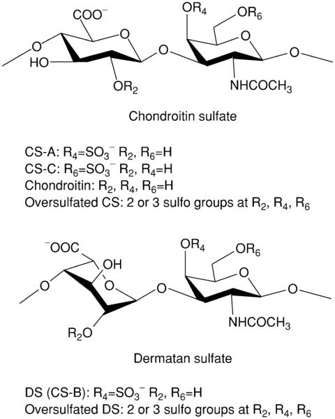
CS, oversulfated CS and chondroitin: the molecular weight ranges from 5000–50,000 (average 25 kDa). DS and oversulfated DS: the molecular weight ranges from 5000–50,000 (average 25 kDa).
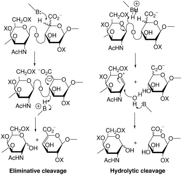
Mechanism for the enzymatic breakdown of GAGs. Lyases catalyze eliminative cleavage and hydrolases catalyze hydrolytic cleavage leading to different oligosaccharide products.
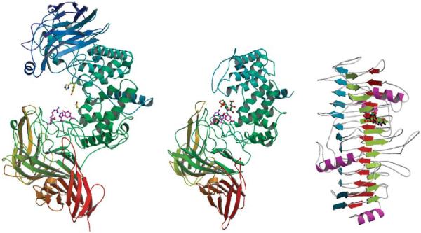
Comparison of crystal structures of chondroitinases PvulABCI (left), FlavoAC (center), and FlavoB (right).
Chondroitin-4,6-sulfate tetrasaccharide in the active site of FlavoAC Tyr234Phe mutant.
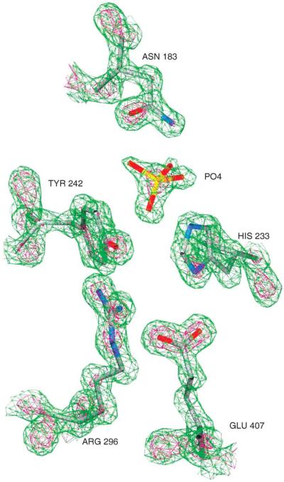
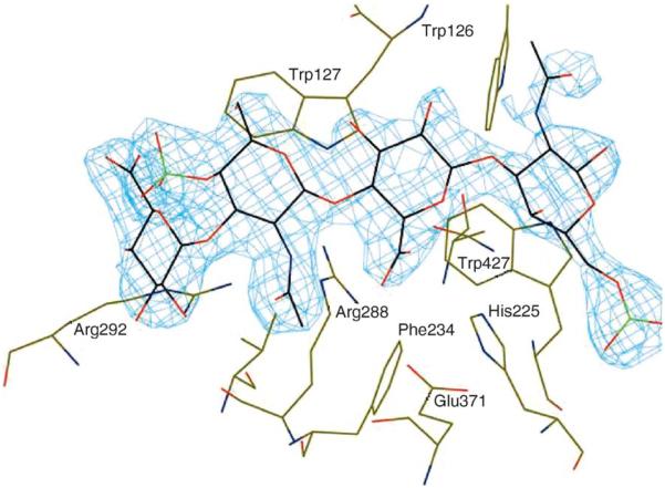
Experimental electron density map of the active site region of ArthroAC. Green contours are drawn at 3σ level, red contours at 5σ level. In the native structure, there is a phosphate ion in the active site. Nitrogen atoms are blue, oxygens are red, carbons are gray, and phosphorus is yellow.
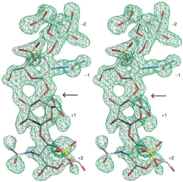
Conformation of the chondroitin-4-sulfate tetrasaccharide substrate bound in the active site of ArthroAC. Omit electron density map is drawn at the 3σ level.
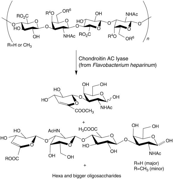
Enzymatic depolymerization of chondroitin O-methyl ester.
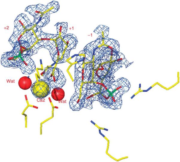
Three disaccharide products bound in the active site of chondroitinase B. Bound calcium atom is shown as a yellow sphere, two water molecules as red spheres.
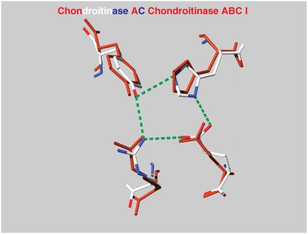
The superposition of the active-site tetrad of FlavoAC and PvulABCI. The Asn175 of FlavoAC and Arg500 of PvulABCI are also shown.
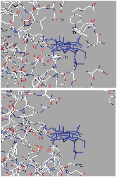
The disposition of the substrate in FlavoAC (left) was transferred to PvulABCI (right) based on the superposition of the active site tetrad. In this open form of PvulABCI are very few contacts between the enzyme and its substrate.
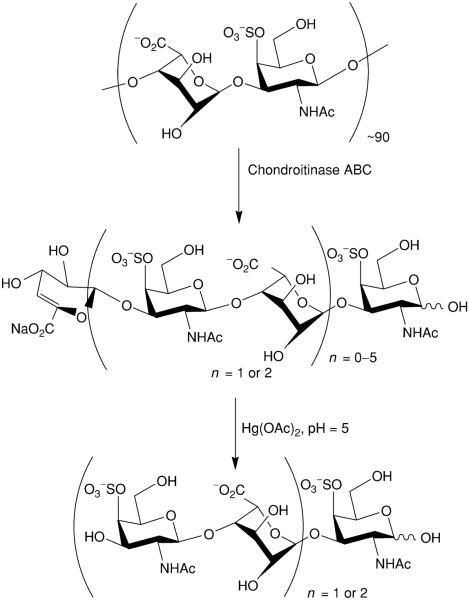
Controlled enzymatic depolymerization of DS by chondroitinase ABC and mercuric acetate treatment to remove the unsaturated nonreducing end residue.
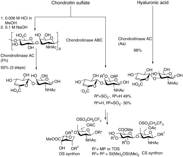
Preparation of desulfated and sulfoprotected disaccharide starting materials for the synthesis of CS/DS/HA oligosaccharides.
Similar articles
-
Degradation of chondroitin sulfate and dermatan sulfate with chondroitin lyases.Methods Mol Biol. 2001;171:363-71. doi: 10.1385/1-59259-209-0:363. Methods Mol Biol. 2001. PMID: 11450250 No abstract available.
-
Chondroitin sulfate lyases: applications in analysis and glycobiology.Adv Pharmacol. 2006;53:167-86. doi: 10.1016/S1054-3589(05)53008-4. Adv Pharmacol. 2006. PMID: 17239766 Review. No abstract available.
-
Identification and biochemical characterization of a novel chondroitin sulfate/dermantan sulfate lyase from Photobacterium sp.Int J Biol Macromol. 2020 Dec 15;165(Pt B):2314-2325. doi: 10.1016/j.ijbiomac.2020.10.119. Epub 2020 Oct 22. Int J Biol Macromol. 2020. PMID: 33132124
-
Facile analysis of contents and compositions of the chondroitin sulfate/dermatan sulfate hybrid chain in shark and ray tissues.Carbohydr Res. 2016 Apr 7;424:54-8. doi: 10.1016/j.carres.2016.02.006. Epub 2016 Feb 24. Carbohydr Res. 2016. PMID: 26986023
-
The structures and applications of microbial chondroitin AC lyase.World J Microbiol Biotechnol. 2022 Aug 23;38(11):199. doi: 10.1007/s11274-022-03395-1. World J Microbiol Biotechnol. 2022. PMID: 35996038 Review.
Cited by
-
Cloning and Characterization of a Chondroitin AC Exolyase from Arthrobacter sp. SD-04.Mol Biotechnol. 2019 Oct;61(10):791-800. doi: 10.1007/s12033-019-00208-z. Mol Biotechnol. 2019. PMID: 31444737
-
Characterization of chondroitin sulfate in stem cells derived from umbilical cord blood in rats.PLoS One. 2022 Jan 25;17(1):e0262854. doi: 10.1371/journal.pone.0262854. eCollection 2022. PLoS One. 2022. PMID: 35077481 Free PMC article.
-
Heterologous production of chondroitin.Biotechnol Rep (Amst). 2022 Feb 10;33:e00710. doi: 10.1016/j.btre.2022.e00710. eCollection 2022 Mar. Biotechnol Rep (Amst). 2022. PMID: 35242620 Free PMC article. Review.
-
A novel eliminase from a marine bacterium that degrades hyaluronan and chondroitin sulfate.J Biol Chem. 2014 Oct 3;289(40):27886-98. doi: 10.1074/jbc.M114.590752. Epub 2014 Aug 13. J Biol Chem. 2014. PMID: 25122756 Free PMC article.
-
Brittlestars contain highly sulfated chondroitin sulfates/dermatan sulfates that promote fibroblast growth factor 2-induced cell signaling.Glycobiology. 2014 Feb;24(2):195-207. doi: 10.1093/glycob/cwt100. Epub 2013 Nov 18. Glycobiology. 2014. PMID: 24253764 Free PMC article.
References
-
- Ahn M, Shin K, Kim D-H, Jang E-A, Toida T, Linhardt R, Kim Y. Characterization of a bacteroides species from human intestine that degrades glycosaminoglycans. Can. J. Microbiol. 1998;44:423–429. - PubMed
-
- Al-Hakim A, Linhardt RJ. Capilary electrophoresis for the analysis of chondroitin sulfate and dermatan sufate derived disaccharides. Anal. Biochem. 1991;195:68–73. - PubMed
-
- Avci FY, Toida T, Linhardt RJ. Chondroitin O-methyl ester: An unusual substrate for chondroitin AC lyase. Carbohydr. Res. 2003;338:2101–2104. - PubMed
-
- Avci FY, Karst N, Islam T, Linhardt RJ. Trifluoroethylsulfonate protected monosaccharides in glycosylation reactions; 228th ACS National Meeting; Philadelphia. 2004.
Publication types
MeSH terms
Substances
Grants and funding
LinkOut - more resources
Full Text Sources
Other Literature Sources
