Comparative Study
. 2007 Apr 1;304(1):246-59.
doi: 10.1016/j.ydbio.2006.12.026. Epub 2006 Dec 15.

Nestin-GFP reporter expression defines the quiescent state of skeletal muscle satellite cells

Affiliations
Comparative Study

Nestin-GFP reporter expression defines the quiescent state of skeletal muscle satellite cells

Kenneth Day et al. Dev Biol. .

Abstract

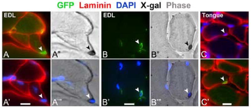
Repair of adult skeletal muscle depends on satellite cells, quiescent myogenic stem cells located beneath the myofiber basal lamina. Satellite cell numbers and performance decline with age and disease, yet the intrinsic molecular changes accompanying these conditions are unknown. We identified expression of GFP driven by regulatory elements of the nestin (NES) gene within mouse satellite cells, which permitted characterization of these cells in their niche. Sorted NES-GFP+ cells exclusively acquired a myogenic fate, even when supplemented with media supporting non-myogenic development. Mutual and unique gene expression by NES-GFP+ cells from hindlimb and diaphragm muscles demonstrated intra- and inter-muscular heterogeneity of satellite cells. NES-GFP expression declined following satellite cell activation and was reacquired in late stage myogenic cultures by non-proliferating Pax7+ progeny. The dynamics of this expression pattern reflect the cycle of satellite cell self-renewal. The NES-GFP model reveals unique transcriptional activity within quiescent satellite cells and permits novel insight into the heterogeneity of their molecular signatures.


Figures

Fig. 1
Fig. 1
GFP Expression within satellite cells in isolated myofibers from NES-GFP/Myf5nlacZ/+ mice. Phase contrast micrographs are merged with images of DAPI-stained nuclei (A–D). Arrows point to individual satellite cells based on GFP expression (A’–D’) and nuclear β-gal staining (A”–B”) or Pax7 immunostaing (C”–D’). Scale bar, 30 μm.
Fig. 2
Fig. 2
Quantification of satellite cells in immunolabeled soleus and EDL myofibers isolated from NES-GFP (A) and NES-GFP/Myf5nlacZ/+ (B) mice. The age of each mouse group is indicated beneath the x-axes. Fibers in the right and left parts of panel B were isolated from the same mice. Each bar depicts the number of satellite cells per individual myofiber. Within each bar, gray segments depict the number of satellite cells that coexpressed Pax7 and GFP (A and B) or GFP and β-gal (B). Green and red segments depict the number of satellite cells that were positive only for GFP, Pax7 or βgal, respectively; β-gal+ only cells were not observed, hence no yellow segments are depicted in panel B.
Fig. 3
Fig. 3
Progeny of satellite cells in EDL myofiber cultures rapidly lose NES-GFP fluorescence upon their activation but regain expression later when entering the reserve state. Phase contrast micrographs are merged with images of DAPI-stained nuclei (A) or BrdU-incorporating nuclei (B). Following one day in culture (A-A”’) only rare Pax7+ satellite cells (red), incorporated BrdU (blue); these rare proliferating cells maintained intense GFP fluorescence like the quiescent (Pax7+/BrdU) satellite cells. GFP fluorescence was drastically reduced by the second culture day as shown for myoblasts still associated with the parent myofiber (B-B”) or emanated from the myofiber (C-C”). These cells expressed MyoD (red) (B”, C”), characteristics of proliferating satellite cells and their progeny. Scale bar, 30 μM (A-A’”), 40 μm (B–C””).
Fig. 4
Fig. 4
Intense NES-GFP expression by satellite cells in cross sections of EDL muscle from NES-GFP/Myf5nlacZ/+ mice (A-A”’, B-B”’) or tongue muscle from NES-GFP mice (C-C’). GFP+ cells (arrowhead) positioned under the myofiber basal lamina as shown by immunolabeling for laminin (red) and DAPI (blue) are also positive for β-gal as shown by merged images of X-gal staining (arrowhead) and phase contrast (A”, B”). Parallel merged images of DAPI stained nuclei and phase images with X-gal staining are also shown (A”’, B”’). Images in panels A and B demonstrate that NES-GFP satellite cells are associated with both small- and large- diameter myofibers of the same muscle Scale bar, 5 μm (A–B”’, C-C’), 10 μm (C-C”’).
Fig. 5
Fig. 5
NES-GFP expression in skeletal muscle microvasculature and satellite cells (AD”) versus Sca1-GFP expression in microvasculature (E–I”). Representative GFP+ capillary networks are shown in partially digested muscles treated with collagenase (EDL) (A, E, F) or Pronase (diaphragm) (B); capillary structure is well-defined in Sca1-GFP muscle under higher magnification (F). NES-GFP+ cells are additionally shown in cross sections of TA muscle (C-C”’ and D-D”); A NES-GFP+ satellite cell (arrowhead) is under the myofiber basal lamina (shown by laminin) (red, converted from far red) and DAPI-stained nucleus (blue) (C-C’), while NES-GFP+ capillary cell (arrow) is outside of the myofiber lamina and colocalizes with the endothelial cell markers CD31 (red) (C”-C”’) and isolectin B4 (red) (D-D”). EDL muscle cross section exhibits Sca1-GFP+ cells between myofibers but not in the satellite cell position under the myofiber basal lamina (G-G’). In representative TA cross sections (H–I”), all Sca1-GFP+ cells coexpress CD31 (red) (H’-H”) and isolectin B4 (red) (I’-I”). Scale bar, 100 μm (A, E), 10 μm (C-C”’), 25 μm (B, D-D”, G-G’, H-H”, I-I”).
Fig. 6
Fig. 6
Scatter plots depicting fluorescence activated cell sorting of cells dissociated from NES-GFP hindlimb muscles. GFP fluorescence plotted against red autofluorescence depicts gating of the most intense GFP positive events (R1) versus negative (R2) (A and B). Sorts from NES-GFP hindlimb yielded 4% GFP events of the total (A), while wildtype hindlimb yielded no GFP fluorescence (B) as shown in the R1 gate. A similar number of negative events were collected for NES-GFP and wildtype muscles as shown by R2 gate (29–32%). Hoechst 33342 plotted against GFP depicts dual Hoechst and GFP-labeled cells in NES-GFP sort, while no GFP fluorescence is detected in the wild-type control as shown by R4 gate (A’ and B’).
Fig. 7
Fig. 7
NES-GFP+ cells from hindlimb muscles are committed to myogenesis. GFP+ and GFP sorted cells were seeded in parallel into myogenic (A, A’ and D), endothelial (B, B’ and E), and neural (C, C’ and F) media, and grown for 1 week. Cultures of GFP+ cells yielded multinucleated myotubes regardless of media type (A-C) while the GFP cells yielded cultures with a non-myogenic phenotype as shown by DAPI and phase contrast overlays (D-F). Double immunostaining for Pax7 (green) and MyoD (red) (A’–C’, merged immunofluorescent images) further demonstrated the uniform myogenic nature of GFP+ progeny cells. Scale bar, 50 μm.
Fig. 8
Fig. 8
RT-PCR analysis of gene expression in satellite cells sorted from hindlimb and diaphragm muscles based on NES-GFP expression. Sorts from Sca1-GFP hindlimb muscle, which served as control for CD31 gene expression, yielded 2.16% GFP events (A). Sorts for NES-GFP hindlimb and diaphragm muscles yielded similar GFP events (Fig. 6). Reactions were performed using cDNA prepared from GFP positive (+) and negative (−) sorted cells, and each reaction included a positive control (con). Cells were prepared from hindlimb muscle of NES-GFP and Sca1-GFP mice (B and D) or NES-GFP diaphragm (C). Total brain RNA was used for each control reaction except for Myf5, MyoD, GFP, and c-met reactions that used total RNA isolated from primary myogenic cultures grown for 5 days.
Fig. 9
Fig. 9
NES-GFP expression is regained in long-term myogenic cultures. Myogenic clones (A–D”) and myofiber cultures (E–F”) were grown to 3 and 4 weeks, respectively. Merged images of GFP fluorescence (green) and immunostaining (red), and parallel merged phase and DAPI stain images are depicted for all antigens examined; unmerged immunostained images are shown for cytoplasmic proteins to discern immunolabel from GFP fluorescence (D, E, and F). All GFP+ cells were positive for Pax7 (A), and typically negative for MyoD except for infrequent MyoD expression in cells expressing only a low level of GFP (B, arrow). GFP+ cells did not express myogenin (C) or saromeric myosin (D and D’). GFP+ cells exhibited polarized localization of nestin compared to GFP (E and E’), and uniform distribution of desmin with GFP (F and F’). Arrowheads in parallel panels point to typical oblong-shaped nuclei found in GFP+ cells. Scale bar, 50 μm (A–D”), 25 μm (E–F”).

References

    1. Aihara M, Sugawara K, Torii S, Hosaka M, Kurihara H, Saito N, Takeuchi T. Angiogenic endothelium-specific nestin expression is enhanced by the first intron of the nestin gene. Lab Invest. 2004;84:1581–1592. - PubMed
    1. Amoh Y, Li L, Katsuoka K, Penman S, Hoffman RM. Multipotent nestin-positive, keratin-negative hair-follicle bulge stem cells can form neurons. Proc Natl Acad Sci U S A. 2005;102:5530–5534. - PMC - PubMed
    1. Banwell BL. Intermediate filament-related myopathies. Pediatr Neurol. 2001;24:257–263. - PubMed
    1. Beauchamp JR, Heslop L, Yu DSW, Tajbakhsh S, Kelly RG, Wernig A, Buckingham ME, Partridge TA, Zammit PS. Expression of CD34 and Myf5 defines the majority of quiescent adult skeletal muscle satellite cells. J Cell Biol. 2000;151:1221–1234. - PMC - PubMed
    1. Brewer GJ, Torricelli JR, Evege EK, Price PJ. Optimized survival of hippocampal neurons in B27-supplemented Neurobasal, a new serum-free medium combination. J Neurosci Res. 1993;35:567–576. - PubMed

Publication types

LinkOut - more resources