. 2007 Feb 6;581(3):437-42.
doi: 10.1016/j.febslet.2006.12.054. Epub 2007 Jan 12.

Tryptophan 334 oxidation in bovine cytochrome c oxidase subunit I involves free radical migration

Affiliations

Tryptophan 334 oxidation in bovine cytochrome c oxidase subunit I involves free radical migration

Patrizia Lemma-Gray et al. FEBS Lett. .

Abstract

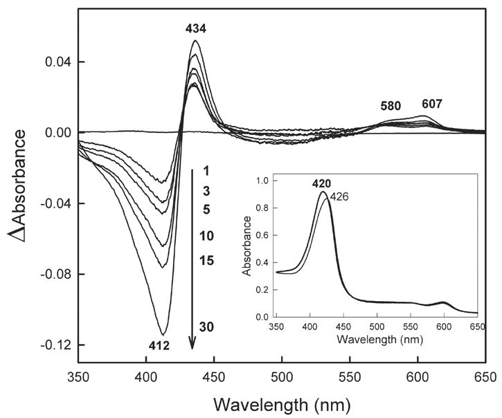
A single tryptophan (W(334(I))) within the mitochondrial-encoded core subunits of cytochrome c oxidase (CcO) is selectively oxidized when hydrogen peroxide reacts with the binuclear center. W(334(I)) is converted to hydroxytryptophan as identified by reversed-phase HPLC-electrospray ionization tandem mass spectrometry analysis of peptides derived from the three SDS-PAGE purified subunits. Total sequence coverage of subunits I, II and III was limited to 84%, 66% and 54%, respectively. W(334(I)) is located on the surface of CcO at the membrane interface. Two other surface tryptophans within nuclear-encoded subunits, W(48(IV)) and W(19(VIIc)), are also oxidized when hydrogen peroxide reacts with the binuclear center (Musatov et al. (2004) Biochemistry 43, 1003-1009). Two aromatic-rich networks of amino acids were identified that link the binuclear center to the three oxidized tryptophans. We propose the following mechanism to explain these results. Electron transfer through the aromatic networks moves the free radicals generated at the binuclear center to the surface-exposed tryptophans, where they produce hydroxytryptophan.


Figures

Figure 1
Figure 1
H2O2-induced changes in the absolute and difference spectrum of cytochrome c oxidase. Main panel: time-dependent difference spectra of H2O2-reacted CcO minus oxidized CcO. Inset: the absolute spectra of oxidized CcO (thick line) and CcO after reaction with 1 mM H2O2 for 30 min. (thin line). Experimental conditions were 6 μM CcO in 20 mM Tris-SO4 buffer at pH 7.4 containing 2 mM dodecylmaltoside.
Figure 2
Figure 2
Total sequence coverage of CcO subunits I, II and III by HPLC-ESI/MS/MS analysis of proteolytic peptides following in-gel digestion of the SDS-PAGE resolved subunits. Chymotryptic peptides detected by MS covered 65%, 46% and 28% of the subunits I, II and III sequences, respectively (sequences are underlined with a simple line). Elastase peptides detected by MS covered 52%, 44% and 38% of the subunit I, II and III sequences, respectively (sequences are underlined with bar-ended line, |——| ). Total sequence coverage by both experimental approaches was 84% of Subunit I, 66% of Subunit II and 54% of Subunit III (sequences are shaded blue). The underlined amino acids indicate the sequence coverage, not proteolytic peptides. For example, chymotryptic coverage of the Subunit I sequence 322–348 was accomplished by MS analysis of chymotryptic peptides 322–327, 328–334, 335–340, 341–344 and 345–348. After exposure of CcO to H2O2, the only oxidized amino acid that could be detected was W334(I) (red box).
Figure 3
Figure 3
Electrospray ionization MS/MS data obtained for chymotryptic peptide 328–334 of Subunit I before (upper panel) and after (lower panel) exposure of CcO to 1 mM H2O2 for 30 min.
Figure 4
Figure 4
Location of the three tryptophans, which are oxidized by hydrogen peroxide, within the 3-dimensional structure of the CcO dimer. The color scheme is: oxidized tryptophans (red), Subunit I (cyan), all other subunits (blue) and the binuclear center (heme a3, gold; CuB, magenta). Image was generated from the 3-dimensional coordinates of CcO (1v54) [25] using PyMol™ software, DeLano Scientific, LLC.
Figure 5
Figure 5
Stereo image of the aromatic amino acid networks within CcO that connect the binuclear center to W334(I), W19(VIIc) and W48(IV). Aromatic amino acids that lead from the binuclear center toward W19(VIIc) are, from left to right: H376(I), H429(I), F430(I), F426(I), F346(I), W323(I), F348(I), Y379(I), F251(I), F344(I), F418(I), F387(I), F414(I), H256(I), W340(I), W396(I), H413(I), H395(I), F78(I), F393(I), F397(I), W409(I), F400(I), F470(I), F476(I), W473(I) and Y403(I). Aromatic amino acids that connect this first network to W334(I) are: Y260(I) and Y261(I). Aromatic amino acids that lead from the binuclear center toward W48(IV) are, from top to bottom: Y372(I), H376(I), H429(I), F430(I), F426(I), F32(II), F321(I), W65(II), F268(I), Y40(II), H328(I), H52(II), Y39(Va), W15(Va), Y18(Va), F68(IV), F19(Va), F64(IV), Y60(IV) and F61(Va). Image was generated from the 3-dimensional coordinates of CcO (1v54) [25] using PyMol™ software, DeLano Scientific, LLC.

References

    1. Cadenas E, Davies KJA. Mitochondrial free radical generation, oxidative stress, and aging. Free Radic Biol Med. 2000;29:222–230. - PubMed
    1. Paradies G, Petrosillo G, Pistolese M, Ruggiero FM. The effect of reactive oxygen species generated from the mitochondrial electron transport chain on the cytochrome c oxidase activity and on the cardiolipin content in bovine heart submitochondrial particles. FEBS Lett. 2000;466:323–326. - PubMed
    1. Turrens JF, Boveris A. Generation of superoxide anion by the NADH dehydrogenase of bovine heart mitochondria. Biochem J. 1980;191:421–427. - PMC - PubMed
    1. Turrens JF, Alexandre A, Lehninger AL. Ubisemiquinone is the electron donor for superoxide formation by Complex III of heart mitochondria. Arch Biochem Biophys. 1985;237:408–414. - PubMed
    1. McLennan HR, Esposti MD. The contribution of mitochondrial respiratory complexes to the production of reactive oxygen species. J Bioenerg Biomembr. 2000;32:153–162. - PubMed

Publication types

MeSH terms