. 2007 May;19(5):1000-10.
doi: 10.1016/j.cellsig.2006.11.004. Epub 2006 Nov 22.

Differential FAK phosphorylation at Ser-910, Ser-843 and Tyr-397 induced by angiotensin II, LPA and EGF in intestinal epithelial cells

Affiliations

Differential FAK phosphorylation at Ser-910, Ser-843 and Tyr-397 induced by angiotensin II, LPA and EGF in intestinal epithelial cells

Xiaohua Jiang et al. Cell Signal. 2007 May.

Abstract

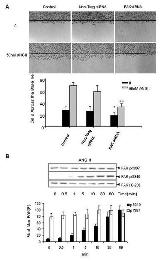
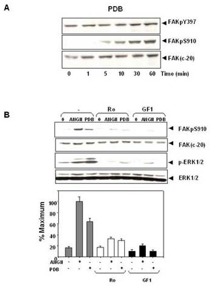
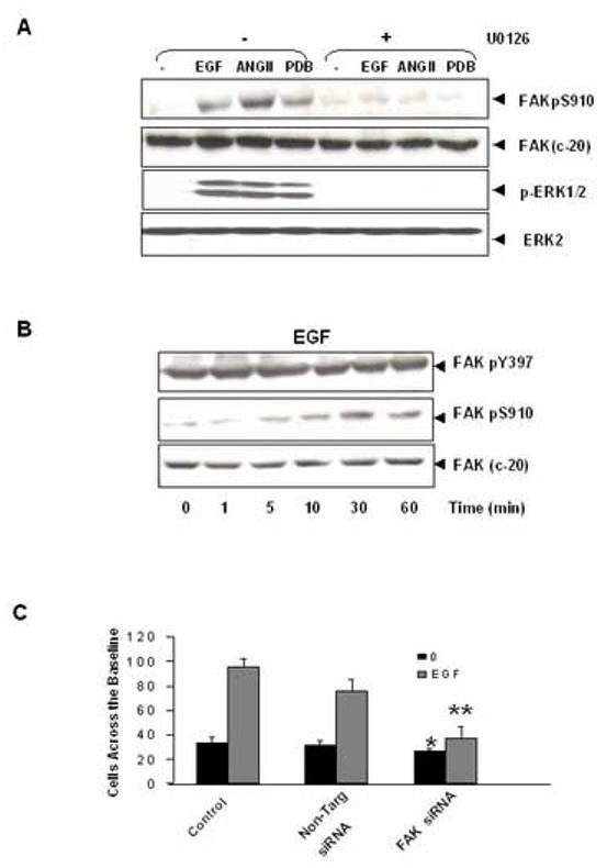
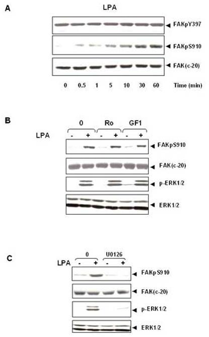
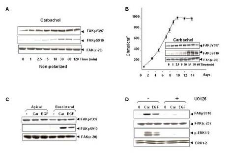
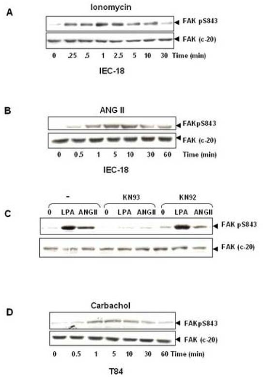
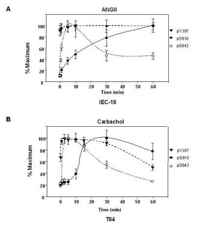
A rapid increase in the tyrosine phosphorylation of the non-receptor tyrosine kinase FAK is a prominent early event in fibroblasts stimulated by a variety of signaling molecules. However, a variety of epithelial cells, including intestinal epithelial cells, show a high basal level of tyrosine phosphorylated FAK that is only slightly further increased by addition of G protein-coupled receptor (GPCR) agonists or growth factors. In this study, we determined whether these stimuli could elicit FAK phosphorylation at serine residues, including Ser-910 and Ser-843. Our results show that multiple agonists including angiotensin II (ANGII), lysophosphatidic acid (LPA), phorbol esters and EGF induced a striking stimulation of FAK phosphorylation at Ser-910 in rat intestinal epithelial IEC-18 cells via an ERK-dependent pathway. In striking contrast, none of these stimuli promoted a significant further increase in FAK phosphorylation at Tyr-397 in these cells. These results were extended using cultures of polarized human colonic epithelial T84 cells. We found that either carbachol or EGF promoted a striking ERK-dependent phosphorylation of FAK at Ser-910, but these agonists caused only slight stimulation of FAK at Tyr-397 in T84 cells. In addition, we demonstrated that GPCR agonists also induced a dramatic increase of FAK phosphorylation at Ser-843 in either IEC-18 or T84 cells. Our results indicate that Ser-910 and Ser-843, rather than Tyr-397, are prominent sites differentially phosphorylated in response to neurotransmitters, bioactive lipids, tumor promoters and growth factors in intestinal epithelial cells.


Figures

Figure 1
Figure 1
ANGII stimulates FAK phosphorylation at Ser-910 in IEC-18 cells. (A). Knockdown FAK expression by siRNA inhibits ANGII induced-cell migration in IEC-18 cells. IEC-18 cells were transfected with Trans IT-TKO alone, non-targeting negative control or 75nM FAK siRNA. After 48 h cells were wounded as described in Materials and Methods and incubated with or without 50nM ANGII for 16h. Experiments were terminated by washing cells twice in PBS, followed by fixing in 10% buffered formalin phosphate for 20 min. Cells were stained with Wright-Giemsa and observed under phase contrast with a ×10 lens mounted on an upright microscope. Upper Panel: Photographs representative of control, non-targeting siRNA or FAKsiRNA transfectd cells. Results are typical of five independent experiments. Lower panel: Values are means ± SE of at least 8 fields from five independent experiments and are expressed as the number of cells observed across the wound margin at 16 h. * P < 0.01 vs. control, ** P<0.01 vs. control with ANGII treatment. (B). Time-course of ANG II in stimulation of FAK phosphorylation at Ser-910 in IEC-18 cells. Upper Panel: Confluent and quiescent IEC-18 cells were treated at 37°C with 50nM of ANG II for various times as indicated and subsequently lysed in 2×sample buffer. FAK phosphorylation at Ser-910 or Tyr-397 was analyzed by Western blotting with pSer-910 Ab or pTyr-397 Ab. The membranes were further analyzed by Western blotting using anti-FAK Ab. The autoradiograms shown are representative of at least three independent experiments. Lower Panel: Quantification of FAK phosphorylation at Ser-910 and Tyr-397 was performed by scanning densitometry. Values shown as bars are the mean of at least three independent experiments and are expressed as the percentage of the maximal increase of phosphorylation above control (unstimulated) values.
Figure2
Figure2
ANG II induces FAK phosphorylation at Ser-910 through a PKC-dependent pathway. (A). Time course of FAK phosphorylation at Ser-910 induced by PDB in IEC-18 cells. Confluent and quiescent cells were treated at 37° C with 100nM PDB for various times as indicated and were subsequently lysed. FAK phosphorylation at Ser-910 or Tyr-397 was analyzed by Western blotting with pSer-910 Ab or pTyr-397 Ab. The membranes were further analyzed by Western blotting using anti-FAK Ab. (B). PKC inhibitors prevent FAK phosphorylation at Ser-910 induced by either ANGII or PDB in IEC-18 cells. Upper Panel: Confluent and quiescent cells were treated for 1 h either in the absence (−) or presence of 2.5 μM Ro-31-8220 or 5μM GF-109203X at 37° C. Cells were then incubated for further 10 min either with 50 nM ANGII or 100 nM PDB. The cells were lysed and the extracts were analyzed by Western blotting with pSer-910 Ab. The membranes were further analyzed by Western blotting using anti-FAK Ab. An aliquot of the lysates was analyzed by Western blotting with phospho-p44/42 MAP Kinase Ab (pERK1/2). These membranes were further analyzed by Western blotting using anti-ERK1/ 2 Ab. Lower Panel: Quantification of the inhibition of Ser-910 phosphorylation by these two PKC inhibitors was performed by scanning densitometry. Values shown as bars are the mean of at least three independent experiments and are expressed as the percentage of the maximal increase of Ser-910 phosphorylation above control (unstimulated) values. In all cases, the autoradiograms shown are representative of at least three independent experiments.
Figure 3
Figure 3
ANGII and EGF induce FAK phosphorylation at Ser-910 through an ERK-dependent pathway. (A). The MEK inhibitor U0126 prevents the phosphorylation of FAK at Ser-910 in IEC-18 cells. Cells were incubated with 10μM U 0126 for 1 h at 37°C and then stimulated with either 5ng/ml EGF or 50 nM ANGII or 100nM PDB for 10 min. The cells were lysed and the extracts were analyzed by Western blotting with pSer-910 Ab. The membranes were further analyzed by Western blotting using anti-FAK Ab. A portion of the lysates was analyzed by Western blotting with pERK1/2 Ab. These membranes were further analyzed by Western blotting using ERK2 Ab. (B). Time-course of EGF in stimulation of FAK phosphorylation at Ser-910 in IEC-18 cells. Confluent and quiescent cells were treated at 37° C with 5 ng/ml EGF for various times as indicated and were subsequently lysed. FAK phosphorylation at Ser-910 or Tyr-397 was analyzed by Western blotting with pSer-910 or pTyr-397. The membranes were further analyzed by Western blotting using anti-FAK Ab. In all cases, the autoradiograms shown are representative of at least three independent experiments. (C). Knockdown FAK expression by siRNA inhibits EGF induced-cell migration in IEC-18 cells. IEC-18 cells were transfected with Trans IT-TKO alone, non-targeting negative control or 75nM FAK siRNA. After 48 h cells were wounded as described in Materials and Methods and incubated with or without 5ng/ml EGF for 16h. Experiments were terminated by washing cells twice in PBS, followed by fixing in 10% buffered formalin phosphate for 20 min. Cells were stained with Wright-Giemsa and observed under phase contrast with a ×10 lens mounted on an upright microscope. Values are means ± SE of at least 8 fields from five independent experiments and are expressed as the number of cells observed across the wound margin at 16 h. * P < 0.01 vs. control, ** P<0.01 vs. control with EGF treatment.
Figure 4
Figure 4
LPA induces FAK phosphorylation at Ser- 910 through PKC-independent, ERK-dependent pathway. (A). Time-course of LPA in stimulation of FAK phosphorylation at Ser-910 in IEC-18 cells. Confluent and quiescent cells were treated at 37° C with 10μM LPA for various times as indicated and were subsequently lysed. FAK phosphorylation at Ser-910 or Tyr-397 was analyzed by Western blotting with pSer-910 Ab or pTyr-397Ab. The membranes were further analyzed by Western blotting using anti-FAK Ab. The autoradiograms shown are representative of at least three independent experiments. (B).LPA stimulates FAK phosphorylation at Ser-910 independent of PKC. Confluent and quiescent cells were treated for 1 h either in the absence (−) or presence of 2.5 μM Ro-31-8220 or 5μM GF-109203X at 37° C. Cells were then incubated for further 10 min with 10μM LPA. The cells were lysed and the extracts were analyzed by Western blotting with pSer-910 Ab. The membranes were further analyzed by Western blotting using anti-FAK Ab. An aliquot of the lysates was analyzed by Western blotting with phospho-p44/42 MAP Kinase Ab (pERK1/2). These membranes were further analyzed by Western blotting using anti-ERK1/ 2 Ab. (C). The MEK inhibitor U0126 prevents LPA induced phosphorylation of FAK at Ser-910 in IEC-18 cells. Cells were incubated with 10μM U 0126 for 1 h at 37°C and then stimulated with 10μM LPA for 10 min. The cells were lysed and the extracts were analyzed by Western blotting with pSer-910 Ab. The membranes were further analyzed by Western blotting using anti-FAK Ab. A portion of the lysates was analyzed by Western blotting with pERK1/2 Ab. These membranes were further analyzed by Western blotting using ERK1/2 Ab. In all cases, the autoradiograms shown are representative of at least three independent experiments.
Figure 5
Figure 5
Carbachol and EGF induce FAK phosphorylation at Ser-910 through ERK-dependent pathway in T84 cells. (A). Time-course of carbachol in stimulation of FAK phosphorylation at Ser-910 and Tyr-397 in un-polarized T84 cells. Cultures of T84 (about 2×10⁶ cells in 35mm Petri dish) were incubated with 100μM carbachol for various times and lysates of these agonist-treated cells were extracted and analyzed by Western blotting using FAK pSer-910 Ab or pTyr-397 Ab. The membranes were further analyzed by Western blotting using anti-FAK Ab. (B). Time-course of carbachol in stimulation of FAK phosphorylation at Ser-910 and Tyr-397 in polarized T84 cells. T84 cells (∼5×10⁵) were plated on 23 mm Falcon Transwells and the transepithelial resistance was measured at various times Inserts, Polarized cultures formed 7 days after plating were stimulated basolaterally with 100μM carbachol for various times. The cells were then lysed and the phosphorylation of FAK at Ser-910 and Tyr-397 was detected by Western blot using FAK pS910Ab or pTyr397 Ab. The same membrane was stripped and reprobed with anti-FAK to verify equal loading. (C). EGF stimulates FAK phosphorylation at Ser-910 in T84 cells. Polarized cultures formed 7 days after plating were stimulated apically or basolaterally with either 100μM carbachol or 50ng/ml EGF for 10 min. The cells were then lysed and the phosphorylation of FAK at Ser-910 or Tyr-397 was detected by Western blot using FAK pSer-910 Ab or pTyr-397 Ab. The same membrane was stripped and reprobed with anti-FAK to verify equal loading. (D). The MEK inhibitor U0126 prevents both carbachol and EGF induced phosphorylation of FAK at Ser-910 in T84 cells. Polarized cultures of cells were incubated with 20μM U 0126 for 1 h at 37°C and then stimulated with either 100μM carbachol or 50ng/ml EGF basolaterally for 10 min. The cells were lysed and the extracts were analyzed by Western blotting with pSer-910 Ab. The membranes were further analyzed by Western blotting using anti-FAK Ab. A portion of the lysates was analyzed by Western blotting with pERK1/2 Ab. These membranes were further analyzed by Western blotting using ERK1/2 Ab. In all cases, the autoradiograms shown are representative of at least three independent experiments.
Figure 6
Figure 6
GPCRs induce FAK phosphorylation at Ser-843 in intestinal epithelial cells. (A). The Ca2+ ionophor ionomycin induces FAK phosphorylation at Ser-843 in IEC-18 cells. Confluent and quiescent IEC-18 cells were treated at 37°C with 500 nM ionomycin for the indicated times. Cells were then lysed and FAK phosphorylation at Ser-843 was analyzed by Western blotting with pSer-843 Ab. The membranes were reanalyzed by Western blotting using anti-FAK Ab (C-20), to demonstrate equal loading of total FAK. The autoradiograms shown are representative of at least three independent experiments. (B). ANGII induces FAK phosphorylation at Ser-843 in IEC-18 cells. Confluent and quiescent IEC-18 cells were treated at 37°C with 50 nM ANGII for the indicated times. Cells were then lysed and analyzed by Western blotting with pSer-843 Ab. The membranes were reanalyzed by Western blotting using anti-FAK Ab, to demonstrate equal loading of total FAK. The autoradiograms shown are representative of at least three independent experiments. (C). CaMKII inhibitor (KN 93) prevents bombesin-induced FAK phosphorylation at Ser-843. Confluent and quiescent IEC-18 cells were preincubated with 40μM KN 93 or KN 92 for 30 min and subsequently stimulated with 50 nM ANGII or 10μM LPA for an additional 1 min. Cells were then lysed and analyzed by Western blotting with pSer-843 Ab. The membranes were reanalyzed by Western blotting using anti-FAK Ab, to demonstrate equal loading of total FAK. The autoradiograms shown are representative of at least three independent experiments. (D). Carbachol induces FAK phosphorylation at Ser-843 in T84 cells. Polarized T84 cells were treated at 37°C with 100μM carbachol basolaterally for the indicated times. Cells were then lysed and analyzed by Western blotting with pSer-843 Ab. The membranes were reanalyzed by Western blotting using anti-FAK Ab, to demonstrate equal loading of total FAK. The autoradiograms shown are representative of at least three independent experiments.
Figure 7
Figure 7
Time-course of GPCRs-induced FAK phosphorylation at Ser-910, Ser-843, and Tyr-397 in intestinal epithelial cells. (A). Time-course of ANGII-induced FAK phosphorylation at Ser-910, Ser-843, and Tyr-397 in IEC-18 cells. Confluent and quiescent IEC-18 cells were treated at 37°C with 50 nM ANGII for various times as indicated in Figs. 1 and 6. Cell lysates were analyzed by Western blotting with pSer-910Ab, pSer-843 Ab and pY-397Ab. Quantification of phosphorylation at Ser-910 (solid line), Tyr-397 (dashed line) and Ser-843 (dotted line) were determined by densitometric scanning of the bands. Values shown are the mean ± S.E. of at least three independent experiments and are expressed as the percentage of the maximal increase in FAK phosphorylation at Ser-910, Tyr-397 or Ser-843 above control (unstimulated) values. (B). Time-course of carbachol-induced FAK phosphorylation at Ser-910, Ser-843, and Tyr-397 in T84 cells. Polarized T84 cells were treated at 37°C with 100μM carbachol basolaterally for various times as indicated in Figs. 5 and 6. Cell lysates were analyzed by Western blotting using pSer-910Ab, pSer-843 Ab and pY-397Ab. Quantification of phosphorylation at Ser-910 (solid line), Tyr-397 (dashed line) and Ser-843 (dotted line) were determined by densitometric scanning of the bands. Values shown are the mean ± S.E. of at least three independent experiments and are expressed as the percentage of the maximal increase in FAK phosphorylation at Ser-910, Tyr-397 or Ser-843 above control (unstimulated) values.

Similar articles

Cited by

References

    1. Zachary I, Rozengurt E. Focal adhesion kinase (p125FAK): a point of convergence in the action of neuropeptides, integrins, and oncogenes. Cell. 1992;71:891–894. - PubMed
    1. Rozengurt E. Convergent signalling in the action of integrins, neuropeptides, growth factors and oncogenes. Cancer Surv. 1995;24:81–96. - PubMed
    1. Hanks SK, Polte TR. Signaling through focal adhesion kinase. Bioessays. 1997;19:137–145. - PubMed
    1. Schlaepfer DD, Hauck CR, Sieg DJ. Signaling through focal adhesion kinase. Prog Biophys Mol Biol. 1999;71:435–478. - PubMed
    1. Schaller MD. Biochemical signals and biological responses elicited by the focal adhesion kinase. Biochim. Biophys. Acta. 2001;1540:1–21. - PubMed

Publication types

MeSH terms