Review
. 2007 Mar;113(3):488-506.
doi: 10.1016/j.pharmthera.2006.11.001. Epub 2006 Nov 28.

Mechanistic pathways and biological roles for receptor-independent activators of G-protein signaling

Affiliations
Review

Mechanistic pathways and biological roles for receptor-independent activators of G-protein signaling

Joe B Blumer et al. Pharmacol Ther. 2007 Mar.

Abstract

Signal processing via heterotrimeric G-proteins in response to cell surface receptors is a central and much investigated aspect of how cells integrate cellular stimuli to produce coordinated biological responses. The system is a target of numerous therapeutic agents and plays an important role in adaptive processes of organs; aberrant processing of signals through these transducing systems is a component of various disease states. In addition to G-protein coupled receptor (GPCR)-mediated activation of G-protein signaling, nature has evolved creative ways to manipulate and utilize the Galphabetagamma heterotrimer or Galpha and Gbetagamma subunits independent of the cell surface receptor stimuli. In such situations, the G-protein subunits (Galpha and Gbetagamma) may actually be complexed with alternative binding partners independent of the typical heterotrimeric Galphabetagamma. Such regulatory accessory proteins include the family of regulator of G-protein signaling (RGS) proteins that accelerate the GTPase activity of Galpha and various entities that influence nucleotide binding properties and/or subunit interaction. The latter group of proteins includes receptor-independent activators of G-protein signaling (AGS) proteins that play surprising roles in signal processing. This review provides an overview of our current knowledge regarding AGS proteins. AGS proteins are indicative of a growing number of accessory proteins that influence signal propagation, facilitate cross talk between various types of signaling pathways, and provide a platform for diverse functions of both the heterotrimeric Galphabetagamma and the individual Galpha and Gbetagamma subunits.


Figures

Figure 1
Figure 1. Properties of AGS proteins in the yeast based functional screen
The details of the yeast-based functional screen are described elsewhere (Cismowski and Lanier, 2005; Cismowski et al., 2002; Cismowski et al., 1999).
Figure 2
Figure 2. Diversity among functional domains in AGS proteins
Schematic diagrams of Group I-III AGS proteins are shown with functional domains highlighted. Domains are as predicted by SMART, PFAM or as described (Takesono et al., 1999). Ras, ras domain - VWA-A, von Willebrand factor A domain - Fn3 - fibronectin type 3 domain, UIM - Ubiquitin-interacting motif, Trip13 - thyroid receptor interacting protein 13, AAA-ATPase - AAA-ATPase domain, TPR – tetratricopeptide repeat. GPR – G-protein regulatory motif.
Figure 3
Figure 3. Functional roles of AGS proteins
Figure 4
Figure 4. Comparison of key domains in Ras and AGS1
Human H-Ras and AGS1 were aligned using the ClustalW program (MacVector™ 7.0) and manual adjustment. A) The three conserved GDP/GTP binding motifs (G1-G3), phosphate-magnesium binding motifs (PM1-PM3) and CAAX motifs were compared. G2 and PM2 regions are identical in the two proteins and are not indicated in this figure. Mutation of amino acids in red result in constitutively active Ras. Residues in Ras that when mutation lead to dominant negative activity are indicated by the single red overlines (S17, D119 in Ras). B) Alignment of the Ras effector domain with the corresponding region of AGS1 and Rhes. Identical residues are indicated by asterisk. This figure is adapted from the thesis of Dr. Govindan Vaidyanathan (2004).
Figure 5
Figure 5. Structure-activity relationships of a consensus GPR peptide for inhibiting GTPγS binding to purified Giα1
A) Identification of the core GPR motif. Amino acid sequence of GPR peptide and truncated GPR peptides. Consensus amino acids are depicted in red. The right column portrays GTPγS binding (500 nM with 2 mM MgCl2) to Giα (100 nM) measured in the absence and presence of GPR peptides (100 μM). B) Sequence alignment and IC50 values for mutated GPR peptides in GTPγS binding assays. Conserved amino acids are depicted in red and mutated amino acids are colored green. Effect of increasing concentrations of competing peptides on GTPγS (500 nM with 2 mM MgCl2) binding to Giα (100 nM). Data are expressed as the percent of specific binding (~1 pmol) observed in the absence of added peptide and are expressed as the mean +/- S.E of two experiments with duplicate determinations. Inactive - inhibition of GTPγS binding was less than 30% at peptide concentrations of 100 μM. Modified from Peterson et al (Peterson et al., 2002).
Figure 6
Figure 6. Accessibility of the GPR-GoLoco binding site on Gαi within the context of heterotrimeric Gαβγ
Left Panel - The model was created by performing a structural carbon-carbon alignment of the Giα subunit in the G protein heterotrimer structure solved by Wall et al (Wall et al., 1995)with the structure of Giα complexed with the GPR-GoLoco peptide derived from RGS14 (Kimple et al., 2002). The Giα subunit from the heterotrimer structure was removed to illustrate how Giα complexed with the GPR-GoLoco peptide would be positioned relative to the Gβ subunit suggesting that that the binding site for peptide binding is accessible in the heterotrimer. Right panel - Comparison of the conformation of Gα switch II in the GPR-Goloco bound (red) or Gβγ bound (green) structures. GiαK210 and GiαE216 are involved in direct electrostatic interactions with Gβγ in the heterotrimer structures.
Figure 7
Figure 7. Schematic of interacting proteins for AGS3 and AGS5/LGN
See Tables 3 and 4 for additional information.
Figure 8
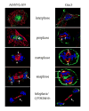
Figure 8. Localization of AGS5/LGN during the cell cycle
CHO cells were synchronized to the entry of mitosis and subsequently allowed to enter the cell cycle and following fixation at increasing times, the cells were stained with antibodies against LGN (green - left panel) or Giα3 (green – right panel) and β-tubulin (red), and DNA stained with DAPI (blue). The yellow signal reflects overlap of the green and red signals. The figure is modified from Blumer et al. 2006.

References

    1. Abba MC, Drake JA, Hawkins KA, Hu Y, Sun H, Notcovich C, Gaddis S, Sahin A, Baggerly K, Aldaz CM. Transcriptomic changes in human breast cancer progression as determined by serial analysis of gene expression. Breast Cancer Res. 2004;6:R499–513. - PMC - PubMed
    1. Adhikari A, Sprang SR. Thermodynamic characterization of the binding of activator of G protein signaling 3 (AGS3) and peptides derived from AGS3 with G alpha i1. J Biol Chem. 2003;278:51825–32. - PubMed
    1. Afshar K, Willard FS, Colombo K, Johnston CA, McCudden CR, Siderovski DP, Gonczy P. RIC-8 is required for GPR-1/2-dependent Galpha function during asymmetric division of C. elegans embryos. Cell. 2004;119:219–30. - PubMed
    1. Bellaiche Y, Gotta M. Heterotrimeric G proteins and regulation of size asymmetry during cell division. Curr Opin Cell Biol. 2005;17:658–63. - PubMed
    1. Bellaiche Y, Radovic A, Woods DF, Hough CD, Parmentier ML, O’Kane CJ, Bryant PJ, Schweisguth F. The Partner of Inscuteable/Discs-large complex is required to establish planar polarity during asymmetric cell division in Drosophila. Cell. 2001;106:355–66. - PubMed

Publication types

MeSH terms

Substances