Comparative Study
. 2007 Jan 30;46(4):1004-12.
doi: 10.1021/bi061774g.

Discovery of two cyanobacterial phenylalanine ammonia lyases: kinetic and structural characterization

Affiliations
Comparative Study

Discovery of two cyanobacterial phenylalanine ammonia lyases: kinetic and structural characterization

Michelle C Moffitt et al. Biochemistry. .

Abstract

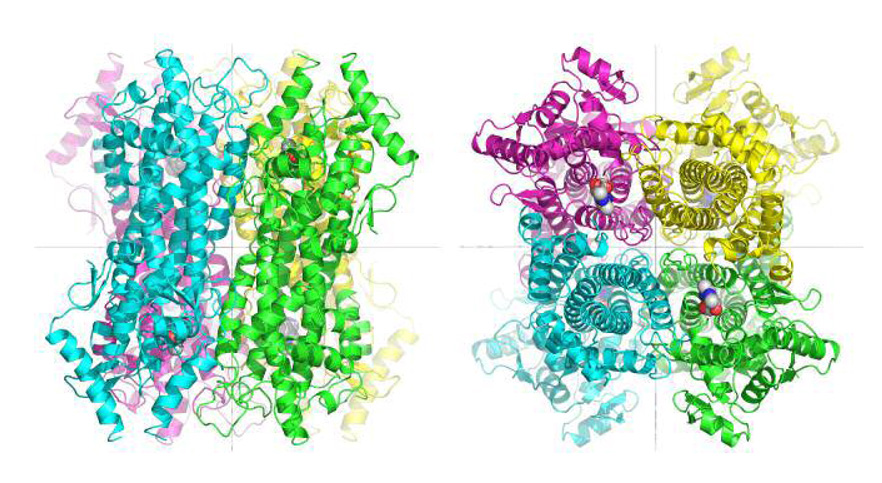
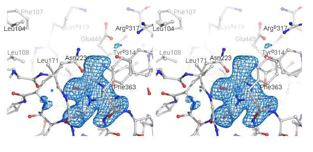
Phenylalanine ammonia lyase (PAL) catalyzes the deamination of phenylalanine to cinnamate and ammonia. While PALs are common in terrestrial plants where they catalyze the first committed step in the formation of phenylpropanoids, only a few prokaryotic PALs have been identified to date. Here we describe for the first time PALs from cyanobacteria, in particular, Anabaena variabilis ATCC 29413 and Nostoc punctiforme ATCC 29133, identified by screening the genome sequences of these organisms for members of the aromatic amino acid ammonia lyase family. Both PAL genes associate with secondary metabolite biosynthetic gene clusters as observed for other eubacterial PAL genes. In comparison to eukaryotic homologues, the cyanobacterial PALs are 20% smaller in size but share similar substrate selectivity and kinetic activity toward L-phenylalanine over L-tyrosine. Structure elucidation by protein X-ray crystallography confirmed that the two cyanobacterial PALs are similar in tertiary and quatenary structure to plant and yeast PALs as well as the mechanistically related histidine ammonia lyases.


Figures

FIGURE 1
FIGURE 1. Structure of cyanobacterial PAL homotetramers and monomers
(A) Ribbon representation of the AvPAL homotetramer, with the polypeptide chains of four individual monomers A (green), B (cyan), C (magenta), and D (yellow). The atoms of the four MIO prosthetic groups are drawn as spheres. The left panel shows a view from the side of the core helical bundles of the four monomers; the right panel shows a top view, into the active-site clefts of two of the monomers. The 222 point-symmetry of the homotetramer is generated by three mutually orthogonal and intersecting two-fold axes (gray lines; two of the axes are visible in each orientation, with the third perpendicular to these two). (B) Stereo ribbon-representation of a monomer of AvPAL. The polypeptide chain is colored according to the colors of the rainbow, with blue for the N-terminus, and red for the C-terminus. Two poorly ordered loops (residues 75 to 91 and 301 to 309) are not included in the structure. The atoms of MIO, formed by the residues Ala167-Ser168-Gly169 of this polypeptide chain are drawn as balls and sticks. The two-fold axes that relate this monomer to the other monomers in the homotetramer are shown as gray lines. This orientation of view here corresponds to a left-side view of the green monomer in Figure 1A. This orientation of view is maintained roughly in all subsequent figures, except for Figure 2.
Figure 2
Figure 2. Comparison of the polypeptide-chain backbones of monomers of AvPAL (green) and parsley PAL (cyan)
Only regions in the vicinity of the inserted shielding domain of parsley PAL (highlighted in darker cyan) are shown. In AvPAL, the shielding domain is replaced by a short loop (dark green).
FIGURE 3
FIGURE 3. Stereo view of the methylidene-imidazolone prosthetic group and protein residues forming the active-site pocket
Protein atoms are drawn as balls and sticks, and are colored by element (carbon: grey; nitrogen: blue; oxygen: red). Hydrogen-bonding interactions formed by MIO are represented as green dashed lines. An oxyanion hole is formed by the backbone amidenitrogens of Leu 172 and Gly 225. The blue-colored contours envelope regions greater than 3σ in the MIO-omit electron-density map (coefficients [Fo-Fc’],ϕc’, where Fc’ and ϕc’ were calculated without contribution from atoms of the MIO).
FIGURE 4
FIGURE 4. Modeled binding of the L-Phe substrate to AvPAL
Hydrogen-bonding interactions formed between the carboxylate group of the L-Phe substrate and the δ-guanido group of AvPAL-Arg 317 are represented as magenta dashed lines. Polypeptide-chain backbones of AvPAL are represented as ribbons, and colored according to monomer as in Figure 3(a). (B) Amino acid differences within the substrate-binding pockets of AvPAL and modeled Streptomyces maritimus are: AvPAL Phe 107 = EncP Ala 82, Leu 108 = Val 83, Lys 419 = Met 399, Ile 423 = Phe 403, Glu 448 = Ala 428.

References

    1. Poppe L, Retey J. Friedel-Crafts-type mechanism for the enzymatic elimination of ammonia from histidine and phenylalanine. Angew Chem Int Ed Engl. 2005;44:3668–3688. - PubMed
    1. Bezanson GS, Desaty D, Emes AV, Vining LC. Biosynthesis of cinnamamide and detection of phenylalanine ammonia-lyase in Streptomyces verticillatus. Can J Microbiol. 1970;16:147–151. - PubMed
    1. Williams JS, Thomas M, Clarke DJ. The gene stlA encodes a phenylalanine ammonia-lyase that is involved in the production of a stilbene antibiotic in Photorhabdus luminescens TT01. Microbiology. 2005;151:2543–2550. - PubMed
    1. Xiang L, Moore BS. Inactivation, complementation, and heterologous expression of encP, a novel bacterial phenylalanine ammonia-lyase gene. J Biol Chem. 2002;277:32505–32509. - PubMed
    1. Berner M, Krug D, Bihlmaier C, Vente A, Muller R, Bechthold A. Genes and enzymes involved in caffeic acid biosynthesis in the actinomycete Saccharothrix espanaensis. J Bacteriol. 2006;188:2666–2673. - PMC - PubMed

Publication types

MeSH terms