. 2007 Mar 1;362(1):44-54.
doi: 10.1016/j.ab.2006.12.023. Epub 2006 Dec 20.

Antibody-based enrichment of peptides on magnetic beads for mass-spectrometry-based quantification of serum biomarkers

Affiliations

Antibody-based enrichment of peptides on magnetic beads for mass-spectrometry-based quantification of serum biomarkers

Jeffrey R Whiteaker et al. Anal Biochem. .

Abstract

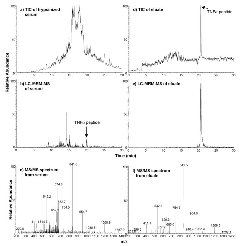
A major bottleneck for validation of new clinical diagnostics is the development of highly sensitive and specific assays for quantifying proteins. We previously described a method, stable isotope standards with capture by antipeptide antibodies, wherein a specific tryptic peptide is selected as a stoichiometric representative of the protein from which it is cleaved, is enriched from biological samples using immobilized antibodies, and is quantitated using mass spectrometry against a spiked internal standard to yield a measure of protein concentration. In this study, we optimized a magnetic-bead-based platform amenable to high-throughput peptide capture and demonstrated that antibody capture followed by mass spectrometry can achieve ion signal enhancements on the order of 10(3), with precision (CVs <10%) and accuracy (relative error approximately 20%) sufficient for quantifying biomarkers in the physiologically relevant ng/mL range. These methods are generally applicable to any protein or biological fluid of interest and hold great potential for providing a desperately needed bridging technology between biomarker discovery and clinical application.


Figures

Figure 1
Figure 1
Optimization of conditions for maximum recovery of captured antigen. (a) Western blot (cropped) showing recovery of 50 ng (2940 fmol) TNFα spiked into 1 mL human serum, captured at 4°C overnight by 20 μL of anti-TNFα polyclonal antibody-coated beads. ‘FT’ refers to flow through. ‘Wash’ refers to 4 times washing of beads with 1 mL of 0.5 M NaCl. Multiple elution buffers were tested, as indicated. Four aliquots of elution buffer (labeled E1–E4, 8 μL each) were added to 20 μL of antigen-bound, antibody-coated beads at RT, in one minute intervals (total of four minutes). At the end of the elution period, the beads were boiled in SDS buffer to remove residual protein (‘Boil’). Note that the secondary antibody used is a goat anti-mouse antibody, which reveals mouse light chain fragments creleased by boiling. (b) SDS-PAGE of antibody enrichment of 50 ng of recombinant TNFα spiked into 400 μL of human serum. The gel image shows recovery of TNFα from a trypsin digest of human serum with (+) and without (−) addition of trypsin inhibitor following the digestion (prior to the antibody-capture step). Addition of trypsin inhibitor results in higher recovery of TNFα protein from trypsin-digested serum.
Figure 2
Figure 2
Calibration curve for quantifying heavy-labeled pure AAC and TNFα peptides. The ion signals for different amounts of pure, synthetic heavy peptides were measured using LC-MS, and used to determine the linear range of quantification on the linear ion trap instrument. Duplicate analyses were performed for each amount of peptide injected. Error bars show the range for each measurement.
Figure 3
Figure 3
Calibration curve for quantifying enriched AAC and TNFα peptides. The ion signal ratio for different amounts of peptides spiked into serum and captured on antibody-coated beads were measured using LC-MS and used to determine the linear range of quantification. For AAC, the ratio of heavy peptide:light peptide (endogeneous) was determined from the extracted ion chromatograms and plotted versus amount of AAC spiked-in. For TNFα, the ratio of light peptide: heavy peptide was determined from the extracted ion chromatograms and plotted versus the expected molar ratio. Three replicate measurements were performed for each target. Error bars show the standard deviation of the measurements. The amounts of the peptides used for the assay characterization studies (Tables 1, 2) were selected at the same points along these curves, within the linear range of the instrument.
Figure 4
Figure 4
Enhancement of endogenous AAC ion signals following antibody enrichment assessed by LC-MS. (a) 30 minute LC-MS analysis of 20 μL depleted and digested human serum. (b) 30 min LC-MRM-MS analysis of AAC from same serum digest. (c) MS/MS spectrum of AAC from serum digest. (d) 30 min LC-MS analysis of antibody-enriched AAC from 20 μL depleted and digested human serum. (e) 30 min LC-MRM-MS analysis of antibody-captured AAC. (f) MS/MS spectrum of AAC from antibody-capture. Equivalent amounts of peptide were injected for LC-MS analysis for each sample (before and after capture).
Figure 5
Figure 5
Enhancement of spiked TNFα ion signals following antibody enrichment assessed by LC-MS. (a) 30 minute LC-MS analysis of 20 μL depleted and digested human serum. (b) 30 min LC-MRM-MS analysis of TNFα from same serum digest. (c) MS/MS spectrum of TNFα from serum digest. (d) 30 min LC-MS analysis of antibody-enriched TNFα from 20 μL depleted and digested human serum. (e) 30 min LC-MRM-MS analysis of antibody-captured TNFα. (f) MS/MS spectrum of TNFα from antibody-capture. Equivalent amounts of peptide were injected for LC-MS analysis for each sample (before and after capture).

References

    1. Clemons M, Danson S, Howell A. Tamoxifen (“Nolvadex”): a review. Cancer Treat Rev. 2002;28:165–80. - PubMed
    1. Haber DA, Bell DW, Sordella R, Kwak EL, Godin-Heymann N, Sharma SV, Lynch TJ, Settleman J. Molecular Targeted Therapy of Lung Cancer: EGFR Mutations and Response to EGFR Inhibitors. Cold Spring Harb Symp Quant Biol. 2005;70:419–26. - PubMed
    1. Ligibel JA, Winer EP. Trastuzumab/chemotherapy combinations in metastatic breast cancer. Semin Oncol. 2002;29:38–43. - PubMed
    1. Shawver LK, Slamon D, Ullrich A. Smart drugs: tyrosine kinase inhibitors in cancer therapy. Cancer Cell. 2002;1:117–23. - PubMed
    1. Fitzgibbons PL, Page DL, Weaver D, Thor AD, Allred DC, Clark GM, Ruby SG, O’Malley F, Simpson JF, Connolly JL, Hayes DF, Edge SB, Lichter A, Schnitt SJ. Prognostic factors in breast cancer. College of American Pathologists Consensus Statement 1999. Arch Pathol Lab Med. 2000;124:966–78. - PubMed

Publication types

LinkOut - more resources