Atypical manifestations of feline inflammatory polyps in three cats
- PMID: 17241805
- PMCID: PMC10822619
- DOI: 10.1016/j.jfms.2006.11.004
Atypical manifestations of feline inflammatory polyps in three cats
Abstract
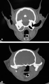
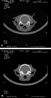
Inflammatory polyps of the feline middle ear and nasopharynx are non-neoplastic masses that are presumed to originate from the epithelial lining of the tympanic bulla or Eustachian tube. The exact origin and cause are unknown, however, it is thought that inflammatory polyps arise as a result of a prolonged inflammatory process. It is unclear whether this inflammation initiates or potentiates the development and growth of inflammatory polyps. Cats with inflammatory polyps typically present with either signs of otitis externa and otitis media or with signs consistent with upper airway obstruction. Traditional diagnostics involve imaging of the tympanic bulla either with skull radiographs or computed topography (CT). Treatment consists of traction and avulsion of the polyp with or without ventral bulla osteotomy (VBO) to remove the epithelial lining of the tympanic bulla. The three cases described here are unusual manifestations or presentations of feline inflammatory polyps that address the following issues: (1) concurrent otic and nasopharyngeal polyps, (2) potential association with chronic viral infection, (3) polyp development in the contralateral middle ear, (4) CT appearance of the skull following VBO, and (5) development of secondary pulmonary hypertension.
Figures


Similar articles
-
Per-endoscopic trans-tympanic traction for the management of feline aural inflammatory polyps: a case review of 37 cats.J Feline Med Surg. 2014 Aug;16(8):645-50. doi: 10.1177/1098612X13516620. Epub 2013 Dec 23. J Feline Med Surg. 2014. PMID: 24366845 Free PMC article.
-
Management of Otic and Nasopharyngeal, and Nasal Polyps in Cats and Dogs.Vet Clin North Am Small Anim Pract. 2016 Jul;46(4):643-61. doi: 10.1016/j.cvsm.2016.01.004. Epub 2016 Mar 4. Vet Clin North Am Small Anim Pract. 2016. PMID: 26947114 Review.
-
Analysis of auditory and neurologic effects associated with ventral bulla osteotomy for removal of inflammatory polyps or nasopharyngeal masses in cats.J Am Vet Med Assoc. 2008 Aug 15;233(4):580-5. doi: 10.2460/javma.233.4.580. J Am Vet Med Assoc. 2008. PMID: 18710312
-
Compartmental location of middle ear inflammatory polyps in cats: 9 cases (2021-2023).J Small Anim Pract. 2025 Mar;66(3):197-202. doi: 10.1111/jsap.13811. Epub 2025 Jan 15. J Small Anim Pract. 2025. PMID: 39814013
-
Feline Aural Inflammatory Polyps.Vet Clin North Am Small Anim Pract. 2025 Mar;55(2):285-298. doi: 10.1016/j.cvsm.2024.11.004. Epub 2025 Jan 16. Vet Clin North Am Small Anim Pract. 2025. PMID: 39824732 Review.
Cited by
-
Megaesophagus in a 6-month-old cat secondary to a nasopharyngeal polyp.J Feline Med Surg. 2010 Apr;12(4):322-4. doi: 10.1016/j.jfms.2009.09.002. J Feline Med Surg. 2010. PMID: 19836983 Free PMC article.
-
Clinical, Radiological, and Echocardiographic Findings in Cats Infected by Aelurostrongylus abstrusus.Pathogens. 2023 Feb 7;12(2):273. doi: 10.3390/pathogens12020273. Pathogens. 2023. PMID: 36839545 Free PMC article.
-
Per-endoscopic trans-tympanic traction for the management of feline aural inflammatory polyps: a case review of 37 cats.J Feline Med Surg. 2014 Aug;16(8):645-50. doi: 10.1177/1098612X13516620. Epub 2013 Dec 23. J Feline Med Surg. 2014. PMID: 24366845 Free PMC article.
-
No evidence of pulmonary hypertension revealed in an echographic evaluation of right-sided hemodynamics in hyperthyroid cats.J Feline Med Surg. 2022 Dec;24(12):e558-e567. doi: 10.1177/1098612X221127102. Epub 2022 Nov 9. J Feline Med Surg. 2022. PMID: 36350661 Free PMC article.
-
Megaesophagus in an 8-month-old cat secondary to a laryngomucocele.JFMS Open Rep. 2024 Jul 25;10(2):20551169241261580. doi: 10.1177/20551169241261580. eCollection 2024 Jul-Dec. JFMS Open Rep. 2024. PMID: 39070187 Free PMC article.
References
-
- Anderson D.M., Robinson R.K., White R.A. Management of inflammatory polyps in 37 cats, The Veterinary Record 147, 2000, 684–687. - PubMed
-
- Blum R.H., McGowan F.X., Jr. Chronic upper airway obstruction and cardiac dysfunction: anatomy, pathophysiology and anesthetic implication, Pediatric Anesthesia 14, 2004, 75–83. - PubMed
-
- Connolly D.J., Lamb C.R., Boswood A. Right-to-left shunting patent ductus arteriosus with pulmonary hypertension in a cat, The Journal of Small Animal Practice 44, 2003, 184–188. - PubMed
-
- Faulkner J.E., Budsberg S.C. Results of ventral bulla osteotomy for the treatment of middle ear polyps in cats, Journal of the American Animal Hospital Association 26, 1990, 496–499.
-
- Herbst L.H., Greiner E.C., Erhart L.M., Bagley D.A., Klein P.A. Serological association between spirorchiadiasis, herpesvirus infection, and fibropapillomatosis in green turtles from Florida, Journal of Wildlife Diseases 34, 1998, 496–507. - PubMed
Publication types
MeSH terms
LinkOut - more resources
Full Text Sources
Medical
Miscellaneous

