. 2007 Jan 29;176(3):307-17.
doi: 10.1083/jcb.200611121. Epub 2007 Jan 22.

SecY alterations that impair membrane protein folding and generate a membrane stress

Affiliations

SecY alterations that impair membrane protein folding and generate a membrane stress

Nobuyuki Shimohata et al. J Cell Biol. .

Abstract

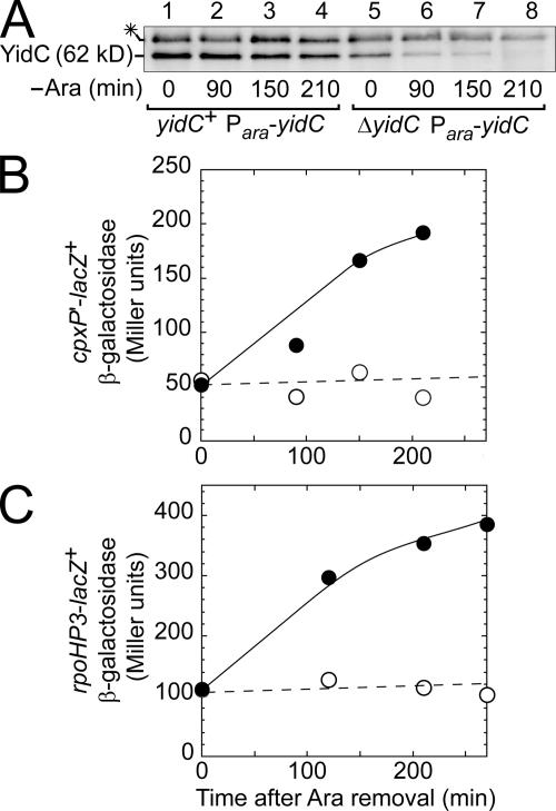
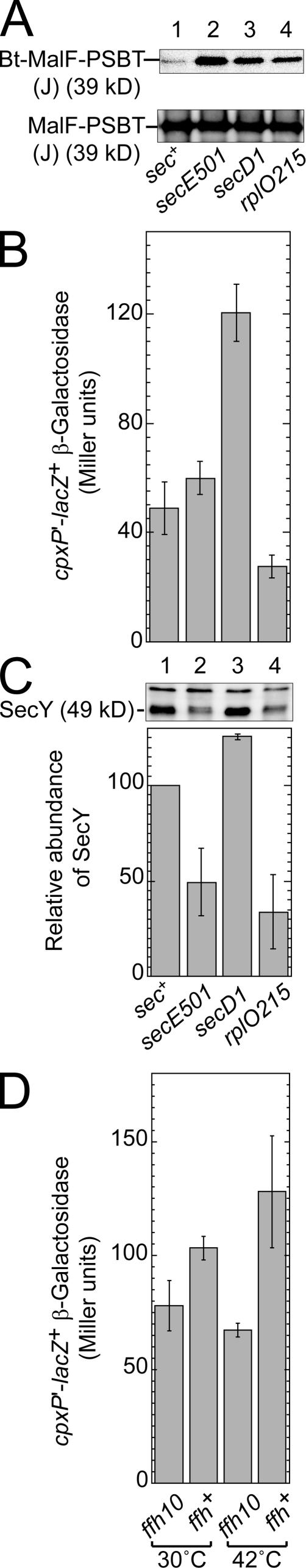
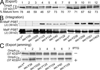
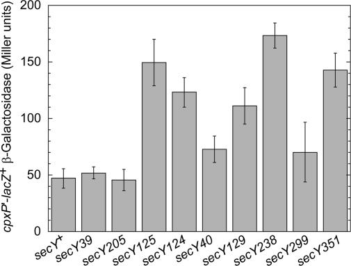
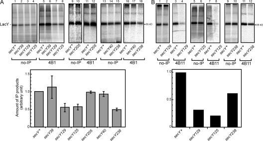
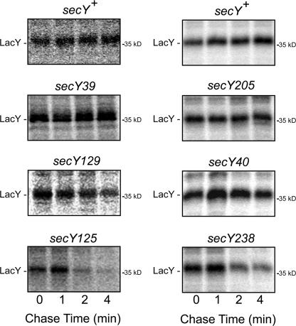
We report on a class of Escherichia coli SecY mutants that impair membrane protein folding. The mutants also up-regulate the Cpx/sigma(E) stress response pathways. Similar stress induction was also observed in response to a YidC defect in membrane protein biogenesis but not in response to the signal recognition particle-targeting defect or in response to a simple reduction in the abundance of the translocon. Together with the previous contention that the Cpx system senses a protein abnormality not only at periplasmic and outer membrane locations but also at the plasma membrane, abnormal states of membrane proteins are postulated to be generated in these secY mutants. In support of this notion, in vitro translation, membrane integration, and folding of LacY reveal that mutant membrane vesicles allow the insertion of LacY but not subsequent folding into a normal conformation recognizable by conformation-specific antibodies. The results demonstrate that normal SecY function is required for the folding of membrane proteins after their insertion into the translocon.


Figures

Figure 1.
Figure 1.
The yidC defect up-regulates extracytoplasmic stress responses. Plasmid pSH10 (cpxP′-lacZ +; A and B) or pSTD643 (rpoHP3-lacZ +; C) was introduced into JS71 (yidC + Para-yidC; A, lanes 1–4; B and C, open circles) and JS7131 (ΔyidC Para-yidC; A, lanes 5–8; B and C, closed circles). The plasmid-bearing cells were grown first in the presence of 0.2% arabinose, washed twice with arabinose-free medium, and grown further in the absence of arabinose for the indicated periods of time. Samples were processed for SDS-PAGE and anti-YidC immunoblotting (A) as well as for β-galactosidase measurements (B and C). The asterisk in A indicates a band of a nonspecific background protein.
Figure 2.
Figure 2.
Stress response induction does not always accompany defects in membrane protein integration. (A) Effects of different sec mutations on MalF-PSBT insertion. Plasmids pSTD343 (lacI q) and pGJ78-J (MalF-PSBT(J)) were introduced into SH463 (sec +), NH192 (secE501), SH625 (secD1), and KI200 (rplO215). The plasmid-bearing cells were grown at 30°C, induced with IPTG for 1 h, and analyzed for biotinylation (top) and accumulation (bottom) of the fusion protein. (B) Effects on Cpx pathway activation. Plasmid pSH10 (cpxP′-lacZ +) was introduced into the sec +, secE501, secD1, and rplO215 strains for β-galactosidase measurements at 30°C. (C) Effects on the SecY contents. Portions of cultures used in B were examined by SDS-PAGE and anti-SecY immunoblotting. The graph shows the cellular contents of SecY relative to the wild-type abundance. (D) The ffh10(Ts) mutation does not activate the Cpx pathway. Plasmid pSH10 (cpxP′-lacZ +) was introduced into SKP1101 (ffh10(Ts)) and SKP1102 (ffh +). The plasmid-bearing cells were grown first at 30°C and then were shifted to 42°C for 1 h for the measurement of β-galactosidase activities. Error bars represent SD.
Figure 3.
Figure 3.
Effects of secY mutations on the export of OmpA and insertion of MalF-PSBT(J). (A) Pulse-labeling analyses of OmpA export. Cells of SH463 (secY +), SH470 (secY39), SH464 (secY205), SH465 (secY125), SH466 (secY124), SH467 (secY40), SH468 (secY129), SH117 (secY +), SH237 (secY238), SH245 (secY299), SH247 (secY351), and SH253 (secY403) were pulse labeled at 37°C with [35S]methionine for 30 s. OmpA was immunoprecipitated and separated by SDS-PAGE into precursor (p) and mature (m) forms. Numbers below each lane show the proportions of radioactivity associated with the mature form after correction for the methionine contents. (B) Biotinylation analyses of MalF-PSBT(J) insertion. Plasmids pSTD343 (lacI q) and pGJ78-J (MalF-PSBT(J) fusion protein) were introduced into SH463 (secY +), SH470 (secY39), SH464 (secY205), SH465 (secY125), SH466 (secY124), SH467 (secY40), SH468 (secY129), SH469 (secY238), SH471 (secY299), SH472 (secY351), and SH253 (secY403). The plasmid-bearing cells were grown at 37°C and induced with IPTG for 1 h. After separation by SDS-PAGE, the MalF fusion proteins were visualized with anti-MalF (bottom), and their biotinylated fractions (Bt-MalF-PSBT(J)) were visualized with streptavidin-conjugated HRP (top). (C) OmpA export in MalF-PSBT(J)–induced cells. The indicated secY mutants, each carrying pSTD343 (lacI q) and pGJ78-J (MalF-PSBT(J)), were grown at 37°C either with (bottom) or without (top) IPTG induction for 1 h. Cells were pulse labeled for 30 s and processed for anti-OmpA immunoprecipitation. Similar results for experiments A–C were obtained at 30°C.
Figure 4.
Figure 4.
Effects of secY mutations on the Cpx stress response. Plasmid pSH10 (cpxP'-lacZ +) was introduced into SH463 (secY +), SH470 (secY39), SH464 (secY205), SH465 (secY125), SH466 (secY124), SH467 (secY40), SH468 (secY129), SH469 (secY238), SH472 (secY299), and SH471 (secY351). Cells were then grown at 30°C, and their β-galactosidase activities were assayed. Similar results were obtained at 37°C. Error bars represent SD.
Figure 5.
Figure 5.
Misfolding of LacY in membranes of secY129, secY238, and secY125. In vitro transcription, translation, and insertion reactions were performed with 4 M urea-washed IMVs prepared from the indicated strains in the presence of 35S-labeled Met and Cys. After the reaction, membranes were collected carefully, subjected to a 4-M urea wash, and solubilized with a detergent. (A) Immunoprecipitation assay with a mAb 4B1. LacY inserted in vitro into IMVs was visualized by SDS-PAGE and autoradiography either directly (lanes 1–4, 9, 10, and 13–15; 5% of the total sample was used) or after immunoprecipitation using mAb 4B1 (lanes 5–8, 11, 12, and 16–18; the rest of the sample was used). It should be noted that the smearing bands at the top regions of the gels also represent the LacY protein, which is known to be prone to aggregation in the presence of SDS. The graph indicates the LacY recovery in the immunoprecipitates as expressed relative to that achieved using wild-type IMV. Data from several independent experiments are averaged, with error bars indicating the SD. IMVs were prepared from TY0 (secY +), AD208 (secY39), TY24 (secY129), and TY8 (secY125) for lanes 1–8 and SH463 (secY +), SH464 (secY205), SH467 (secY40), and SH469 (secY238) for lanes 9–18. (B) Immunoprecipitation assay with mAb 4B11. The experiment was performed as in A using mAb 4B11 for immunoprecipitation as indicated. IMVs were prepared from TY0 (secY +), TY24 (secY129), TY8 (secY125), and AD2397 (secY238).
Figure 6.
Figure 6.
LacY is destabilized in membrane protein folding–defective secY mutants. LacY-His10 was expressed in isogenic strains TY0 (secY +), AD208 (secY39), TY24 (secY129), and TY8 (secY125) in the left panels and SH463 (secY +), SH464 (secY205), SH467 (secY40), and SH469 (secY238) in the right panels as described in Materials and methods. Cells were pulse labeled for 1 min with radioactive [35S]methionine and cysteine and were chased with excess amounts of nonradioactive methionine and cysteine for the indicated time periods. Membranes were isolated from the labeled cells and washed with urea. LacY was isolated from the urea-washed membranes by means of metal affinity chromatography, subjected to SDS-PAGE, and autoradiographed.

References

    1. Abramson, J., I. Smirnova, V. Kasho, G. Verner, H.R. Kaback, and S. Iwata. 2003. Structure and mechanism of the lactose permease of Escherichia coli. Science. 301:610–615. - PubMed
    1. Akiyama, Y., and K. Ito. 1989. Export of Escherichia coli alkaline phosphatase attached to an integral membrane protein, SecY. J. Biol. Chem. 264:437–442. - PubMed
    1. Akiyama, Y., and K. Ito. 1990. SecY protein, a membrane-embedded secretion factor of E. coli, is cleaved by the OmpT protease in vitro. Biochem. Biophys. Res. Commun. 167:711–715. - PubMed
    1. Akiyama, Y., T. Inada, Y. Nakamura, and K. Ito. 1990. SecY, a multispanning integral membrane protein, contains a potential leader peptidase cleavage site. J. Bacteriol. 172:2888–2893. - PMC - PubMed
    1. Akiyama, Y., K. Kanehara, and K. Ito. 2004. RseP (YaeL), an E. coli RIP protease, cleaves transmembrane sequences. EMBO J. 23:4434–4442. - PMC - PubMed

Publication types

MeSH terms