. 2007 Feb 1;21(3):278-91.
doi: 10.1101/gad.1498707. Epub 2007 Jan 22.

The enhancement of pericentromeric cohesin association by conserved kinetochore components promotes high-fidelity chromosome segregation and is sensitive to microtubule-based tension

Affiliations

The enhancement of pericentromeric cohesin association by conserved kinetochore components promotes high-fidelity chromosome segregation and is sensitive to microtubule-based tension

Carrie A Eckert et al. Genes Dev. .

Abstract

Sister chromatid cohesion, conferred by the evolutionarily conserved cohesin complex, is essential for proper chromosome segregation. Cohesin binds to discrete sites along chromosome arms, and is especially enriched surrounding centromeres, but past studies have not clearly defined the roles of arm and pericentromeric cohesion in chromosome segregation. To address this issue, we developed a technique that specifically reduced pericentromeric cohesin association on a single chromosome without affecting arm cohesin binding. Under these conditions, we observed more extensive stretching of centromeric chromatin and elevated frequencies of chromosome loss, suggesting that pericentromeric cohesin enrichment is essential for high-fidelity chromosome transmission. The magnitude of pericentromeric cohesin association was negatively correlated with tension between sister kinetochores, with the highest levels of association in cells lacking kinetochore-microtubule attachments. Pericentromeric cohesin recruitment required evolutionarily conserved components of the inner and central kinetochore. Together, these observations suggest that pericentromeric cohesin levels reflect the balance of opposing forces: the kinetochore-mediated enhancement of cohesin binding and the disruption of binding by mechanical tension at kinetochores. The involvement of conserved kinetochore components suggests that this pathway for pericentromeric cohesin enrichment may have been retained in higher eukaryotes to promote chromosome biorientation and accurate sister chromatid segregation.


Figures

Figure 1.
Figure 1.
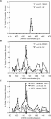
Pericentromeric cohesin enrichment is prevented by centromere-flanking “insulators.” Wild-type (CEY70) and I-CEN3-I (CEY40) cells containing Mcd1-6HA were released from G1 arrest into media containing 15 μg/mL NZ. Cultures were processed for ChIP when >90% large budded, indicating metaphase arrest. ChIP was performed using HA antiserum. DNA cross-linked to Mcd1 and diluted input DNA not subjected to immunoprecipitation were analyzed by PCR using primer pairs that amplify ∼300-bp fragments within the CHRIII pericentromeric region (A) or at a centromere-distal CHRIII region (B). Arm and pericentromeric Mcd1 binding data are plotted on equivalent scales for comparison. Quantitation of DNA in the Mcd1 ChIPs, expressed as a percentage of the input DNA, is plotted as a function of the locations of the midpoints of those DNA fragments based on the Saccharomyces Genome Database (SGD) coordinates. The position of the centromere is indicated by a black oval.
Figure 2.
Figure 2.
Disruption of pericentromeric cohesin enrichment extends the domain subject to stretching. LacOp arrays were placed to the left of CEN3, ∼19 kb away on wild-type (CEY90) or ∼23 kb away in I-CEN3-I (CEY91) cells (at SGD coordinate 95678) that express a LacI-GFP fusion protein. Cells were staged in G1 with α-factor and released in fresh medium lacking pheromone. Aliquots of cells were collected for an asynchronous control, G1-arrested cells, and at 15-min intervals post-release, and fixed in paraformaldehyde. Bud morphology and the number and position of GFP spots were scored for each time point (n ≥ 100).
Figure 3.
Figure 3.
Disruption of pericentromeric cohesin enrichment decreases chromosome transmission fidelity. (A) A schematic of the MATa I-CEN3-I/MATα wild-type CEN3 diploid is shown. CEN3 is indicated by the oval, and the G + C-rich Myxococcus DNA and the PGK1 region flanking the centromere are shown as black and white boxes, respectively. URA3 is integrated near the telomere on the MATa homolog. (B) Chromosome loss was determined using quantitative mating assays (Materials and Methods). A haploid MATα strain and diploids that were either monosomic for MATα CHRIII (CEY102) or contained a MATa copy marked with URA3 for selection and either wild-type (WT) CEN3 (CEY39) or I-CEN3-I (CEY61) were crossed to MATa and MATα haploid testers to determine their ability to mate. Mating due to loss of the CHRIII MATa homolog, as opposed to gene conversion of the MAT locus, was determined by growth on minimal media lacking uracil.
Figure 4.
Figure 4.
Mcd1p binding in the CEN3 pericentromeric region increases in the absence of microtubules and is independent of the spindle assembly checkpoint. (A) Mcd1-6HA cdc16 cells (1829-15B) were released from G1 arrest into 37°C prewarmed media containing either 15 μg/mL NZ or DMSO until arrested (∼3 h), as determined by large-budded cell morphologies. Cells were processed for ChIP using HA antibody. DNA cross-linked to Mcd1 and diluted input DNA were analyzed as described in Figure 1 for a CHRXII arm region (A) and the CHRIII pericentromeric region (B). (C) Mcd1-6HA pGAL-MPS1 cells (PMY319) were staged in G1 in rich media containing raffinose (2% final concentration). Cells were released into NZ- or DMSO-containing media in the presence or absence of galactose (4% final concentration) to induce Mps1p overexpression. Cells were processed for ChIP when mostly large budded, and cell samples were collected for flow-cytometric analyses to confirm metaphase enrichment (data not shown). The quantitation of CHRIII pericentromeric DNA in Mcd1 ChIPs was done as described in the legend for Figure 1.
Figure 5.
Figure 5.
Kinetochore mutants that alter microtubule attachments and/or tension exhibit increased pericentromeric Mcd1p levels. Mutant and isogenic controls were staged in G1 using α-factor and then released into NZ- or DMSO-containing 37°C prewarmed rich media. Cells were processed for ChIP using antibodies against epitope-tagged Mcd1 (Mcd1-6HA or Mcd1-18MYC) when mostly large budded. Metaphase enrichment of cells was confirmed by flow-cytometric analyses (data not shown). Quantitation of CHRIII pericentromeric DNA in Mcd1 ChIPs was performed as described in the legend for Figure 1. (A) Results for Mcd1-18MYC cdc16-123 ipl1-321 (1701-38C) or IPL1 (1700-55A) are shown. (B) Results for Mcd1-6HA cdc16-1 ndc80-1 (1849-40D) or NDC80 (1847-22C) are shown. (C) Results for Mcd1-6HA stu2-277 (1704-19B) or STU2 (1704-18B) are shown.
Figure 6.
Figure 6.
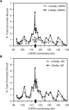
Monopolar attachment results in increased pericentromeric Mcd1p levels. Mcd1-6HA cdc6Δ pGAL-Ubi-R-CDC6 cells (1705-6A) were grown in rich medium with raffinose and galactose (2% each) to allow expression of CDC6. Cells were staged in G1 and released into galactose-containing medium for 20 min to allow replication to initiate. The culture was then split into two, and one half was washed and resuspended in glucose-containing medium to repress CDC6 expression, while the other half was maintained in galactose-containing medium. Each culture was again treated with α-factor to arrest cells at the beginning of the next cell cycle. After G1 arrest, both cultures were released in either 15 μg/mL NZ- or DMSO-containing medium without further change in the medium’s carbon source. Cells were collected when mostly large budded, and DNA contents of each culture was confirmed by flow-cytometric analyses (data not shown). (A) The levels of CHRIII pericentromeric sequences in Mcd1 ChIPs in DMSO alone control cells are shown. (B) The levels of CHRIII pericentromeric sequences in Mcd1 ChIPs in NZ-treated cells are shown.
Figure 7.
Figure 7.
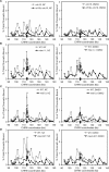
Components of the central and inner kinetochore are required for pericentromeric cohesin enrichment. (A) Mcd1-6HA cdc16-123 ctf19Δ (CEY57) or isogenic CTF19 (1829-15B) cells were staged in G1 and released into 37°C prewarmed media containing either NZ or DMSO. Cells were processed for Mcd1 ChIP when mostly large budded. Cell samples were taken immediately preceding formaldehyde cross-linking for flow-cytometric analyses to confirm metaphase enrichment (data not shown). Analysis of ChIP DNA was done as described in Figure 1. (B) Mcd1-6HA mif2-3 (CEY68) or isogenic MIF2 (CEY67) cells were treated as described in A and processed for Mcd1 ChIP. (C) Mcd1-6HA mtw1-1 (CEY47) or isogenic MTW1 (CEY46) cells were treated as described in A and processed for Mcd1 ChIP. (D) Mcd1-6HA cse4-327 (CEY49) or isogenic CSE4 (CEY46) cells were grown to saturation, diluted into fresh medium, and grown at 37°C for 20 min before addition of NZ or DMSO.

Comment in

References

    1. Allshire R.C., Nimmo E.R., Ekwall K., Javerzat J.P., Cranston G., Nimmo E.R., Ekwall K., Javerzat J.P., Cranston G., Ekwall K., Javerzat J.P., Cranston G., Javerzat J.P., Cranston G., Cranston G. Mutations derepressing silent centromeric domains in fission yeast disrupt chromosome segregation. Genes & Dev. 1995;9:218–233. - PubMed
    1. Amor D.J., Choo K.H., Choo K.H. Neocentromeres: Role in human disease, evolution, and centromere study. Am. J. Hum. Genet. 2002;71:695–714. - PMC - PubMed
    1. Bernard P., Maure J.F., Partridge J.F., Genier S., Javerzat J.P., Allshire R.C., Maure J.F., Partridge J.F., Genier S., Javerzat J.P., Allshire R.C., Partridge J.F., Genier S., Javerzat J.P., Allshire R.C., Genier S., Javerzat J.P., Allshire R.C., Javerzat J.P., Allshire R.C., Allshire R.C. Requirement of heterochromatin for cohesion at centromeres. Science. 2001;294:2539–2542. - PubMed
    1. Biggins S., Murray A.W., Murray A.W. The budding yeast protein kinase Ipl1/Aurora allows the absence of tension to activate the spindle checkpoint. Genes & Dev. 2001;15:3118–3129. - PMC - PubMed
    1. Biggins S., Severin F.F., Bhalla N., Sassoon I., Hyman A.A., Murray A.W., Severin F.F., Bhalla N., Sassoon I., Hyman A.A., Murray A.W., Bhalla N., Sassoon I., Hyman A.A., Murray A.W., Sassoon I., Hyman A.A., Murray A.W., Hyman A.A., Murray A.W., Murray A.W. The conserved protein kinase Ipl1 regulates microtubule binding to kinetochores in budding yeast. Genes & Dev. 1999;13:532–544. - PMC - PubMed

Publication types

Substances

LinkOut - more resources