. 2007 Apr;27(7):2466-75.
doi: 10.1128/MCB.01641-06. Epub 2007 Jan 22.

Swapping the gene-specific and regional silencing specificities of the Hst1 and Sir2 histone deacetylases

Affiliations

Swapping the gene-specific and regional silencing specificities of the Hst1 and Sir2 histone deacetylases

Janet Mead et al. Mol Cell Biol. 2007 Apr.

Abstract

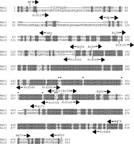
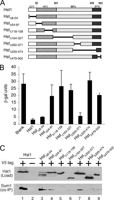
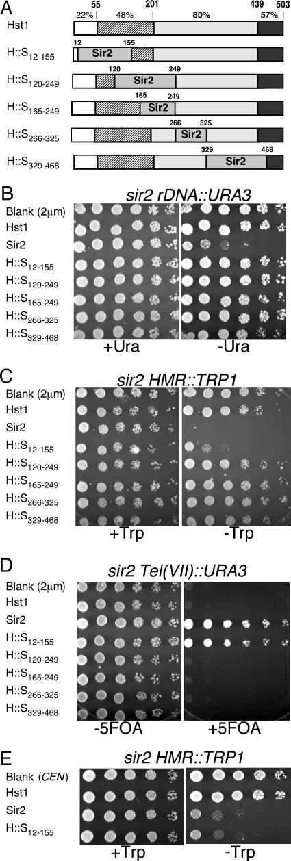
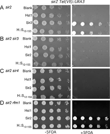
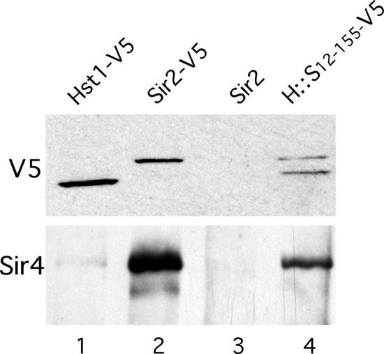
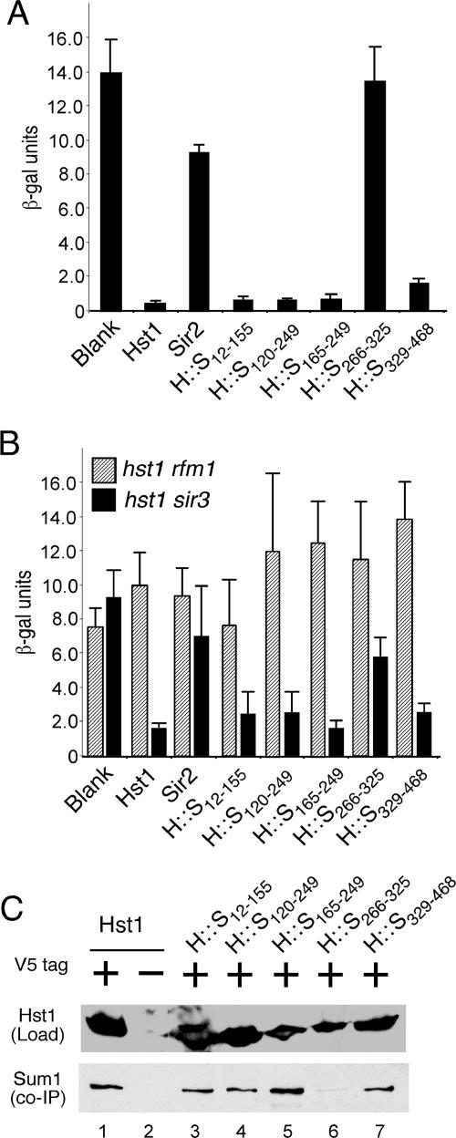
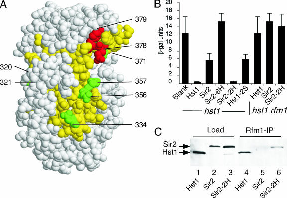
Sir2 and Hst1 are NAD(+)-dependent histone deacetylases of budding yeast that are related by strong sequence similarity. Nevertheless, the two proteins promote two mechanistically distinct forms of gene repression. Hst1 interacts with Rfm1 and Sum1 to repress the transcription of specific middle-sporulation genes. Sir2 interacts with Sir3 and Sir4 to silence genes contained within the silent-mating-type loci and telomere chromosomal regions. To identify the determinants of gene-specific versus regional repression, we created a series of Hst1::Sir2 hybrids. Our analysis yielded two dual-specificity chimeras that were able to perform both regional and gene-specific repression. Regional silencing by the chimeras required Sir3 and Sir4, whereas gene-specific repression required Rfm1 and Sum1. Our findings demonstrate that the nonconserved N-terminal region and two amino acids within the enzymatic core domain account for cofactor specificity and proper targeting of these proteins. These results suggest that the differences in the silencing and repression functions of Sir2 and Hst1 may not be due to differences in enzymatic activities of the proteins but rather may be the result of distinct cofactor specificities.


Figures

FIG. 1.
FIG. 1.
Sequence similarity of Sir2 and Hst1. Amino acid alignment of the yeast Sir2 and Hst1 proteins. Identical residues are shaded in gray. Endpoints of the internal deletions constructed in Hst1 are shown with arrows above the sequence. Junctions of the Hst1::Sir2 chimeras indicating the Hst1 residues that were replaced with the corresponding regions of Sir2 are shown with arrows below the sequence. The asterisks indicate positions in which residues in Sir2 were replaced with the amino acids at the corresponding positions in Hst1.
FIG. 2.
FIG. 2.
Deletion analysis of Hst1. (A) Schematic of the deletions that were constructed in Hst1. The top line shows the conservation of different domains of Hst1 with Sir2. The numbers above the line indicate the endpoints of the different regions of the protein, and the percentages are the sequence identity with Sir2. Each mutant was named for the residues that were deleted. (B) MSE repression assays of wild-type and Hst1 deletions. The indicated mutants on high-copy-number 2μm plasmids were tested for the ability to repress transcription of an MSE-regulated promoter driving lacZ expression in JXY5, an hst1Δ strain. The bars indicate the average β-galactosidase activities of five independent isolates, and the standard deviations are shown by the error bars. (C) (Top) Western blot with antibody to the V5 epitope to monitor the levels of expression of tagged (lane 1) and untagged (lane 2) wild-type Hst1 or the indicated V5-tagged Hst1 deletion mutants (lanes 3 to 9). (Bottom) Western blot with Myc antibody to detect the presence of Myc-tagged Sum1 protein that coimmunoprecipitated with tagged or untagged wild-type Hst1 or the indicated mutants.
FIG. 3.
FIG. 3.
Transcriptional silencing by Hst1::Sir2 chimeras. (A) A schematic representation of the Hst1::Sir2 chimeras is shown. The top line shows a schematic of Hst1 with numbers indicating the percent identity of the different regions of the protein with Sir2. The rows below show the different Hst1::Sir2 chimeras, in which the numbers indicate the positions of the residues in Hst1 that were replaced with the corresponding residues in Sir2. (B) High-copy-number (2μm) plasmids containing either wild-type SIR2, HST1, or the indicated HST1::SIR2 chimeras were transformed into strain LPY2447 (sir2 rDNA::URA3), and normalized serial fivefold dilutions of the cultures were spotted on plates with (+Ura) and without (−Ura) uracil. Failure to grow on −Ura medium indicates silencing of the rDNA loci. (C) Wild-type and chimera constructs were transformed in strain LPY3923 (sir2 hmr::TRP1), and normalized serial dilutions of the cultures were spotted on media with (+Trp) and without (−Trp) tryptophan. Failure to grow on −Trp medium indicates silencing of the HMR loci. (D) Normalized cultures of the indicated transformants of strain LPY1953 (sir2 TEL::URA3) were serially diluted and spotted on media with and without the drug 5-FOA. 5-FOA prevents growth of the cells that express URA3. The ability to grow on the 5-FOA plate indicates silencing of the telomeric loci in the cell. (E) Low-copy-number (CEN) plasmids containing either wild-type SIR2, HST1, or the HST1::SIR212-155 chimera were transformed into strain LPY3923 and assayed as for panel C.
FIG. 4.
FIG. 4.
Silencing by the Hat1::Sir212-155 chimera is dependent on Sir3 and Sir4 but independent of Rfm1. Plasmids containing wild-type HST1 or SIR2 or the Hat1::Sir212-155 chimera, along with a blank vector, were transformed in strain (A) LPY1953 (sir2 TEL::URA3), (B) JMY049 (LPY1953 with sir3Δ::KanMX), (C) JMY053 (LPY1953 with sir4Δ::KanMX), and (D) JMY058 (LPY1953 with rfm1Δ::KanMX), and normalized fivefold serial dilutions of cultures were spotted on plates with and without 5-FOA. Growth on 5-FOA indicates silencing of the telomere loci.
FIG. 5.
FIG. 5.
The Hat1::Sir212-155 chimera coimmunoprecipitates with Sir4. Lysates from transformants of strain JXY20 (sir2Δ hst1Δ) containing plasmids with V5-tagged wild-type Hst1 (lane 1), V5-tagged Sir2 (lane 2), untagged Sir2 (lane 3), or V5-tagged Hat1::Sir212-155 chimera (lane 4) were immunoprecipitated with antibody to the V5 epitope. The top panel shows a Western blot with antibody to the V5 epitope to monitor the levels of expression of V5-tagged proteins in crude lysates. The bottom panel shows a Western blot of the immunoprecipitated pellets using polyclonal antibodies to Sir4.
FIG. 6.
FIG. 6.
MSE-mediated repression by the Hst1::Sir2 chimeras. (A) Plasmids containing either wild-type SIR2, HST1, or the indicated HST1::SIR2 chimeras were transformed into strain JXY5 (hst1Δ::KanMX) and assayed for the ability to repress the MSE-regulated promoter on plasmid pJX43. The bars indicate the average levels of β-galactosidase activity of five independent transformants, and the standard deviations are shown by the error bars. (B) Wild-type SIR2, HST1, or the indicated HST1::SIR2 chimeras were transformed into strain RMY8 (hst1Δ::KanMX rfm1Δ::KanMX) (hatched bars) or JMY045 (hst1Δ::KanMX sir3Δ::HIS3) (solid bars) and assayed for the ability to repress the MSE-regulated promoter as in panel A. (C) The upper blot shows a Western blot analysis with antibody to the V5 epitope to compare the levels of expression of tagged (lane 1) and untagged (lane 2) wild-type Hst1 and the indicated Hst1::Sir2 chimeras (lanes 3 to 7). Extracts from these strains were immunoprecipitated with antibodies specific for the V5 epitope, and the pellets were assayed by Western blot analysis with Myc antibodies to detect the presence of Myc epitope-tagged Sum1 in the immunoprecipitate (bottom).
FIG. 7.
FIG. 7.
Residues in the 266-to-325 region of Hst1 specify interactions with Rfm1. (A) A space-filling model of the human SIRT2 protein that was derived from the crystal structure is shown (12). This view is of the back side of the protein, away from the NAD+ binding pocket and the predicted binding site of the histone tail. The region corresponding to Hst1 residues 266 to 325 (Sir2 residues 314 to 379) is shown in yellow. The residues in green are the positions in the Sir2-6H mutant that were replaced with the corresponding amino acids in Hst1. The residues in red correspond to positions N378 and L379 in Sir2 that were replaced in the Sir2-2H mutant. (B) The level of repression of an MSE-regulated promoter by Hst1, Sir2, Sir2-6H (K320N, I321M, M334D, S356D, T357P, and T371S), Sir2-2H (N378Q and L379I), and Hst1-2S (Q324N and I325L) mutants on low-copy-number CEN plasmids were assayed as described in the legend to Fig. 2B. The assays for the six bars on the left were performed in strain JXY5 (hst1Δ), while the assays for the three bars on the right were performed in strain RMY8 (hst1Δ rfm1Δ). (C) A co-IP assay showed that Sir2-2H interacts with Rfm1. Strain JXY5 (hst1Δ::KanMX) was cotransformed with high-copy-number 2μm plasmids expressing RFM1-HA and HST1 (lanes 1 and 4), SIR2 (lanes 2 and 5), and SIR2-2H (lanes 3 and 6). A Western blot of the crude lysates (lanes 1 to 3) and Rfm1-immunoprecipitated samples (lanes 4 to 6) was probed with polyclonal antibody to the V5 epitope.

Similar articles

Cited by

References

    1. Ai, W., P. G. Bertram, C. K. Tsang, T. F. Chan, and X. F. Zheng. 2002. Regulation of subtelomeric silencing during stress response. Mol. Cell. 10:1295-1305. - PubMed
    1. Avalos, J. L., I. Celic, S. Muhammad, M. S. Cosgrove, J. D. Boeke, and C. Wolberger. 2002. Structure of a Sir2 enzyme bound to an acetylated p53 peptide. Mol. Cell 10:523-535. - PubMed
    1. Brachmann, C. B., J. M. Sherman, S. E. Devine, E. E. Cameron, L. Pillus, and J. D. Boeke. 1995. The SIR2 gene family, conserved from bacteria to humans, functions in silencing, cell cycle progression, and chromosome stability. Genes Dev. 9:2888-2902. - PubMed
    1. Chang, J. H., H. C. Kim, K. Y. Hwang, J. W. Lee, S. P. Jackson, S. D. Bell, and Y. Cho. 2002. Structural basis for the NAD-dependent deacetylase mechanism of Sir2. J. Biol. Chem. 277:34489-34498. - PubMed
    1. Cockell, M. M., S. Perrod, and S. M. Gasser. 2000. Analysis of Sir2p domains required for rDNA and telomeric silencing in Saccharomyces cerevisiae. Genetics 154:1069-1083. - PMC - PubMed

Publication types

MeSH terms

LinkOut - more resources