. 2007 Mar;27(6):2059-73.
doi: 10.1128/MCB.01828-06. Epub 2007 Jan 22.

The Myc transactivation domain promotes global phosphorylation of the RNA polymerase II carboxy-terminal domain independently of direct DNA binding

Affiliations

The Myc transactivation domain promotes global phosphorylation of the RNA polymerase II carboxy-terminal domain independently of direct DNA binding

Victoria H Cowling et al. Mol Cell Biol. 2007 Mar.

Abstract

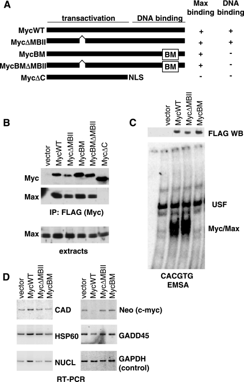
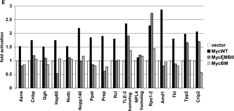
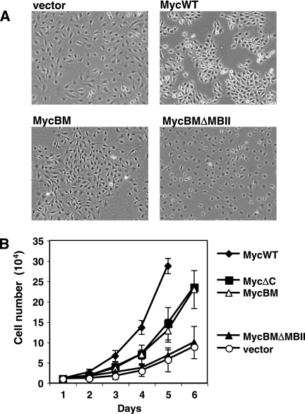
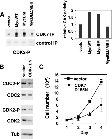
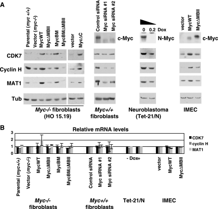
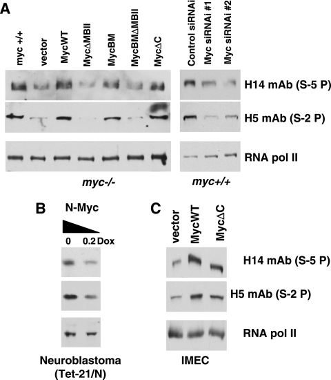
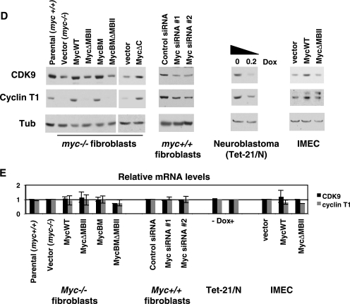
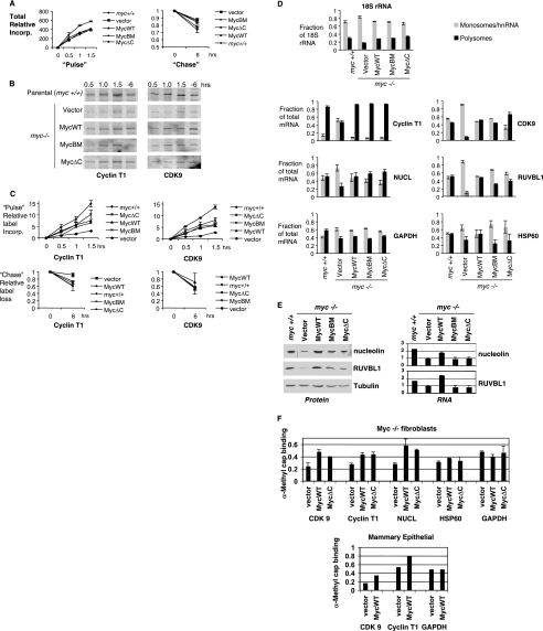
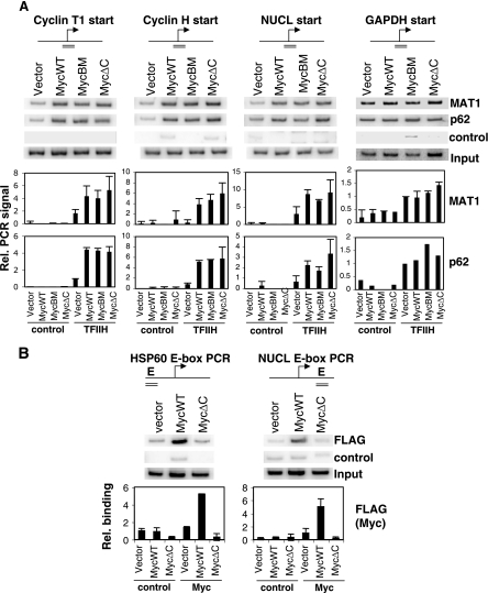
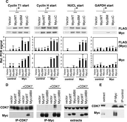
Myc is a transcription factor which is dependent on its DNA binding domain for transcriptional regulation of target genes. Here, we report the surprising finding that Myc mutants devoid of direct DNA binding activity and Myc target gene regulation can rescue a substantial fraction of the growth defect in myc(-/-) fibroblasts. Expression of the Myc transactivation domain alone induces a transcription-independent elevation of the RNA polymerase II (Pol II) C-terminal domain (CTD) kinases cyclin-dependent kinase 7 (CDK7) and CDK9 and a global increase in CTD phosphorylation. The Myc transactivation domain binds to the transcription initiation sites of these promoters and stimulates TFIIH binding in an MBII-dependent manner. Expression of the Myc transactivation domain increases CDK mRNA cap methylation, polysome loading, and the rate of translation. We find that some traditional Myc transcriptional target genes are also regulated by this Myc-driven translation mechanism. We propose that Myc transactivation domain-driven RNA Pol II CTD phosphorylation has broad effects on both transcription and mRNA metabolism.


Figures

FIG. 1.
FIG. 1.
DNA binding-deficient Myc mutants. (A) A diagram of murine N-Myc mutants. MycΔMBII has a deletion of amino acids 103 to 119. MycBM has amino acids 381 to 384 mutated from RQRR to ADAA. MycBMΔMBII has both previous mutations. MycΔC is truncated at amino acid 370 and has a nuclear localization signal (NLS) at the C terminus. DNA binding in EMSA and Max binding in coimmunoprecipitation are summarized. (B) Myc proteins were immunoprecipitated from reconstituted myc−/− cells using anti-FLAG antibody and immunoblotted for N-Myc and Max (upper panels). Cell extracts were immunoblotted for Max (lower panel). (C) EMSA was performed on extracts from 293 cells expressing the indicated Myc proteins, using a Myc/Max consensus binding site probe. Extracts were also immunoblotted with anti-FLAG to detect the Myc proteins (upper panel). (D) RNA extracted from myc−/− cell lines expressing the indicated Myc protein was used for RT-PCR using primers specific for the Myc-activated CAD, HSP60, and nucleolin (NUCL) genes, the Myc-repressed neomycin (Neo) (under the control of the c-myc promoter) and GADD45 genes, and GAPDH as a control. (E) The same RNA was hybridized to rat oligonucleotide microarrays (see Materials and Methods). The graph shows the relative expression levels of Myc target genes in response to MycWT, MycΔMBII, and MycBM compared to that of the vector control. When the expression levels of the samples were compared to those of the vector control, the t test returned the following P values: MycWT, 1.4 × 10⁻⁷; MycΔMBII, 0.09; and MycBM, 0.5. WB, Western blot; USF, upstream stimulatory factor.
FIG. 1.
FIG. 1.
DNA binding-deficient Myc mutants. (A) A diagram of murine N-Myc mutants. MycΔMBII has a deletion of amino acids 103 to 119. MycBM has amino acids 381 to 384 mutated from RQRR to ADAA. MycBMΔMBII has both previous mutations. MycΔC is truncated at amino acid 370 and has a nuclear localization signal (NLS) at the C terminus. DNA binding in EMSA and Max binding in coimmunoprecipitation are summarized. (B) Myc proteins were immunoprecipitated from reconstituted myc−/− cells using anti-FLAG antibody and immunoblotted for N-Myc and Max (upper panels). Cell extracts were immunoblotted for Max (lower panel). (C) EMSA was performed on extracts from 293 cells expressing the indicated Myc proteins, using a Myc/Max consensus binding site probe. Extracts were also immunoblotted with anti-FLAG to detect the Myc proteins (upper panel). (D) RNA extracted from myc−/− cell lines expressing the indicated Myc protein was used for RT-PCR using primers specific for the Myc-activated CAD, HSP60, and nucleolin (NUCL) genes, the Myc-repressed neomycin (Neo) (under the control of the c-myc promoter) and GADD45 genes, and GAPDH as a control. (E) The same RNA was hybridized to rat oligonucleotide microarrays (see Materials and Methods). The graph shows the relative expression levels of Myc target genes in response to MycWT, MycΔMBII, and MycBM compared to that of the vector control. When the expression levels of the samples were compared to those of the vector control, the t test returned the following P values: MycWT, 1.4 × 10⁻⁷; MycΔMBII, 0.09; and MycBM, 0.5. WB, Western blot; USF, upstream stimulatory factor.
FIG. 2.
FIG. 2.
DNA binding-deficient Myc mutants induce cell proliferation and morphological change. The results of an analysis of myc−/− cells expressing MycWT and mutants are shown. (A) Phase-contrast micrographs of log-phase cells. (B) Growth curves. The data shown represent the means for at least three experiments, and error bars show the standard deviations.
FIG. 3.
FIG. 3.
The DNA binding-deficient Myc mutant elevates total cellular CAK activity. (A) A CAK assay was performed on extracts from myc−/− fibroblasts expressing the indicated Myc protein. Protein immunoprecipitated with anti-CDK7 or control antibody was incubated with recombinant CDK2 and [32P]ATP, and reaction products were run on a gel, visualized by a phosphorimager (left panel), and quantitated (right panel). The results of a representative experiment are shown. (B) Cell extracts were prepared from myc+/+ fibroblasts (TGR) expressing the vector control and CDK7 DN as indicated. Extracts were immunoblotted using antibodies raised against phospho-CDC2 (CDC2-P), CDC2, phospho-CDK2 (CDK2-P), CDK2, and γ-tubulin (Tub). (C) Growth curves for myc+/+ fibroblasts (TGR) expressing the vector control and CDK7 DN. The data shown represent the means for two experiments, and error bars show the standard deviations.
FIG. 4.
FIG. 4.
Myc induces CAK expression by a posttranscriptional mechanism. (A) Cell extracts were prepared from myc+/+ or myc−/− fibroblasts expressing the indicated Myc protein from several cell systems: myc+/+ fibroblasts transfected for 24 h with control siRNA or two independent Myc siRNAs, a neuroblastoma cell line (Tet-21/N) expressing doxycycline-off N-Myc cultured in 0 and 0.2 μg/ml doxycycline (Dox) for 2 days, and IMECs expressing c-MycWT or c-MycΔMBII. Immunoblot analyses were performed on cell extracts using antibodies raised against CAK components, CDK7, cyclin H, and MAT1, and γ-tubulin (Tub). Immunoblot analyses were also performed using anti-c-Myc antibodies on myc+/+ cell extracts and IMEC extracts and using anti-N-Myc antibodies on neuroblastoma cell extracts. (B) RNA was extracted from two independent log-phase samples. RT-PCR was performed in the linear range using primers specific for CDK7, cyclin H, MAT1, and GAPDH. Mean relative expression levels were calculated, and error bars indicate the standard deviations.
FIG. 5.
FIG. 5.
Myc induces RNA Pol II phosphorylation. Nuclear extracts were prepared from the following log-phase cells: (A) myc+/+ fibroblasts, myc−/− fibroblasts expressing the indicated Myc protein, myc+/+ fibroblasts transfected with control siRNA or two independent Myc siRNAs, (B) a neuroblastoma cell line (Tet-21/N) expressing N-Myc cultured in 0 and 0.2 μg/ml doxycycline (Dox) for 2 days, and (C) IMECs expressing the indicated Myc protein. Immunoblot analyses were performed using monoclonal antibodies (mAb) raised against RNA Pol II CTD phospho-S5 (H14) and phospho-S2 (H5) and the RNA Pol II large subunit. (D) Immunoblot analyses were performed on cell extracts using antibodies raised against P-TEFb components, CDK9 and cyclin T1, and γ-tubulin (Tub). (E) RT-PCR was performed in the linear range using primers specific for CDK9 and cyclin T1 on RNA from two independent samples. Mean relative expression levels were calculated, and error bars indicate the standard deviations.
FIG. 5.
FIG. 5.
Myc induces RNA Pol II phosphorylation. Nuclear extracts were prepared from the following log-phase cells: (A) myc+/+ fibroblasts, myc−/− fibroblasts expressing the indicated Myc protein, myc+/+ fibroblasts transfected with control siRNA or two independent Myc siRNAs, (B) a neuroblastoma cell line (Tet-21/N) expressing N-Myc cultured in 0 and 0.2 μg/ml doxycycline (Dox) for 2 days, and (C) IMECs expressing the indicated Myc protein. Immunoblot analyses were performed using monoclonal antibodies (mAb) raised against RNA Pol II CTD phospho-S5 (H14) and phospho-S2 (H5) and the RNA Pol II large subunit. (D) Immunoblot analyses were performed on cell extracts using antibodies raised against P-TEFb components, CDK9 and cyclin T1, and γ-tubulin (Tub). (E) RT-PCR was performed in the linear range using primers specific for CDK9 and cyclin T1 on RNA from two independent samples. Mean relative expression levels were calculated, and error bars indicate the standard deviations.
FIG. 6.
FIG. 6.
Myc elevates cyclin T1 and CDK9 translation rate, polysome loading, and mRNA cap methylation. Log-phase myc+/+ fibroblasts and myc−/− fibroblasts expressing MycWT, MycBM, MycΔC, or the vector control were labeled (“pulse”) with [35S]methionine/cysteine for 0.5, 1.0, or 1.5 h. Subsequently, cells were washed and incubated in regular growth medium (“chase”) for 6 h. Cell extracts were prepared and normalized for protein content. (A) Protein was precipitated from cell extracts using TCA and the counts detected using a scintillation counter. The left panel represents label incorporation (Incorp.) during “pulse,” and the right panel represents label loss during “chase.” (B) Cyclin T1 and CDK9 were immunoprecipitated from cell extracts, resolved on SDS-PAGE, and visualized by a phosphorimager. (C) Mean quantitation of duplicates from a representative experiment is shown. Error bars show the standard deviations. The upper panels indicate relative label incorporation into cyclin T1 and CDK9 during “pulse,” and the lower panels indicate label loss during “chase.” (D) Using the same cell lines as described above, polysomes were separated from monosomes/hnRNA by centrifugation through a sucrose bed. The proportion of 18S rRNA in the polysome fraction and monosome fraction was calculated. mRNA from each fraction was used as substrate for RT-PCR for the mRNA indicated. The proportion of each mRNA in polysome and monosome fractions for each cell line is depicted. The results of a representative experiment are shown. Mean values of duplicates are shown, and error bars indicate the standard deviations. (E) Protein extracts made from the same panel of cell lines were used to perform Western blot analyses, probing with anti-RUVBL1 and anti-nucleolin (NUCL) antibodies. RT-PCR was performed on independently isolated RNA samples from the same cells by using primers specific for RUVBL1 and nucleolin. (F) Cap methylation was assessed for RNA extracted from myc−/− fibroblasts expressing the vector control, MycWT, and MycΔC and from immortalized mammary epithelial cells expressing the vector control and MycWT. RNA was subjected to immunoprecipitation using anti-2,2,7-methylguanosine antibody. RNA was purified and used as a template for RT-PCR using primers specific for the genes indicated. The quantity of immunoprecipitated RNA is expressed as a fraction of input RNA. The results of representative experiments are shown.
FIG. 7.
FIG. 7.
Myc promotes TFIIH binding and binds to the transcription initiation sites via the N terminus. ChIP was performed on myc−/− fibroblasts expressing MycWT, MycBM, MycΔC, or the vector control. (A) Complexes were immunoprecipitated with antibodies raised against the TFIIH subunits MAT1 and p62 or control antibody. Coprecipitated DNA was used as a template for PCR using primers specific for cyclin T1, cyclin H, nucleolin (NUCL), and GAPDH transcriptional start sites. PCR products were labeled, run on a gel, and visualized by a phosphorimager (upper panels). PCR products were quantitated and normalized to input PCR, and the relative (Rel.) signal is depicted. The mean values for at least two independent experiments are shown in the graphs. Error bars show the standard deviations. (B) Complexes were immunoprecipitated with anti-FLAG (Myc) or control antibody. PCR was carried out using primers specific for HSP60 and nucleolin E boxes. (C) Complexes were immunoprecipitated with antibodies raised against the FLAG (Myc) tag and Myc or control antibody. PCR was carried out using primers specific for cyclin T1, cyclin H, nucleolin, and GAPDH transcription start sites. (D) 293 cells were transfected with MycWT, MycΔMBII, and CDK7 as indicated. Immunoprecipitation was carried out with cell extracts using anti-CDK7 (left panel) or anti-Myc (middle panel). Immunoprecipitated proteins and cell extracts (right panel) were subjected to Western blotting using anti-CDK7 and anti-Myc antibodies. (E) Immunoprecipitation was carried out using anti-c-Myc antibodies or control antibodies on untransfected 293 cells. Immunoprecipitated protein and cell extracts were subjected to Western blotting using anti-CDK7 and anti-Myc antibodies.
FIG. 7.
FIG. 7.
Myc promotes TFIIH binding and binds to the transcription initiation sites via the N terminus. ChIP was performed on myc−/− fibroblasts expressing MycWT, MycBM, MycΔC, or the vector control. (A) Complexes were immunoprecipitated with antibodies raised against the TFIIH subunits MAT1 and p62 or control antibody. Coprecipitated DNA was used as a template for PCR using primers specific for cyclin T1, cyclin H, nucleolin (NUCL), and GAPDH transcriptional start sites. PCR products were labeled, run on a gel, and visualized by a phosphorimager (upper panels). PCR products were quantitated and normalized to input PCR, and the relative (Rel.) signal is depicted. The mean values for at least two independent experiments are shown in the graphs. Error bars show the standard deviations. (B) Complexes were immunoprecipitated with anti-FLAG (Myc) or control antibody. PCR was carried out using primers specific for HSP60 and nucleolin E boxes. (C) Complexes were immunoprecipitated with antibodies raised against the FLAG (Myc) tag and Myc or control antibody. PCR was carried out using primers specific for cyclin T1, cyclin H, nucleolin, and GAPDH transcription start sites. (D) 293 cells were transfected with MycWT, MycΔMBII, and CDK7 as indicated. Immunoprecipitation was carried out with cell extracts using anti-CDK7 (left panel) or anti-Myc (middle panel). Immunoprecipitated proteins and cell extracts (right panel) were subjected to Western blotting using anti-CDK7 and anti-Myc antibodies. (E) Immunoprecipitation was carried out using anti-c-Myc antibodies or control antibodies on untransfected 293 cells. Immunoprecipitated protein and cell extracts were subjected to Western blotting using anti-CDK7 and anti-Myc antibodies.

Similar articles

Cited by

References

    1. Adhikary, S., F. Marinoni, A. Hock, E. Hulleman, N. Popov, R. Beier, S. Bernard, M. Quarto, M. Capra, S. Goettig, U. Kogel, M. Scheffner, K. Helin, and M. Eilers. 2005. The ubiquitin ligase HectH9 regulates transcriptional activation by Myc and is essential for tumor cell proliferation. Cell 123:409-421. - PubMed
    1. Amati, B., T. D. Littlewood, G. I. Evan, and H. Land. 1993. The c-Myc protein induces cell cycle progression and apoptosis through dimerization with Max. EMBO J. 12:5083-5087. - PMC - PubMed
    1. Arabi, A., S. Wu, K. Ridderstrale, H. Bierhoff, C. Shiue, K. Fatyol, S. Fahlen, P. Hydbring, O. Soderberg, I. Grummt, L. G. Larsson, and A. P. Wright. 2005. c-Myc associates with ribosomal DNA and activates RNA polymerase I transcription. Nat. Cell Biol. 7:303-310. - PubMed
    1. Armelin, H. A., M. C. Armelin, K. Kelly, T. Stewart, P. Leder, B. H. Cochran, and C. D. Stiles. 1984. Functional role for c-myc in mitogenic response to platelet-derived growth factor. Nature 10:55-660. - PubMed
    1. Bentley, D. L. 2005. Rules of engagement: co-transcriptional recruitment of pre-mRNA processing factors. Curr. Opin. Cell Biol. 17:251-256. - PubMed

Publication types

MeSH terms

LinkOut - more resources