. 2007 Mar;13(3):317-27.
doi: 10.1261/rna.220407. Epub 2007 Jan 22.

The role of the S1 domain in exoribonucleolytic activity: substrate specificity and multimerization

Affiliations

The role of the S1 domain in exoribonucleolytic activity: substrate specificity and multimerization

Mónica Amblar et al. RNA. 2007 Mar.

Abstract

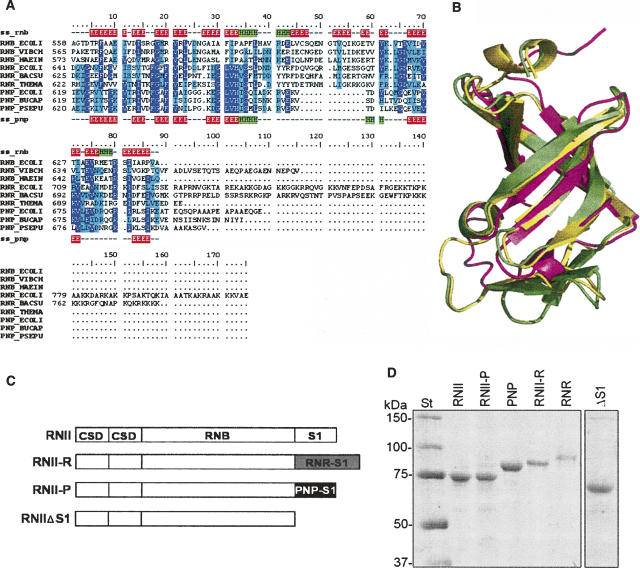
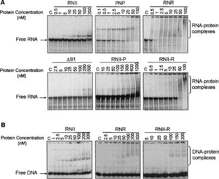
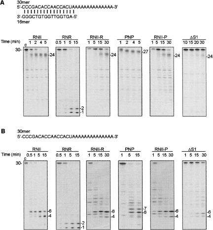
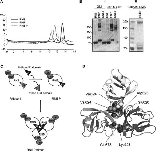
RNase II is a 3'-5' exoribonuclease that processively hydrolyzes single-stranded RNA generating 5' mononucleotides. This enzyme contains a catalytic core that is surrounded by three RNA-binding domains. At its C terminus, there is a typical S1 domain that has been shown to be critical for RNA binding. The S1 domain is also present in the other major 3'-5' exoribonucleases from Escherichia coli: RNase R and polynucleotide phosphorylase (PNPase). In this report, we examined the involvement of the S1 domain in the different abilities of these three enzymes to overcome RNA secondary structures during degradation. Hybrid proteins were constructed by replacing the S1 domain of RNase II for the S1 from RNase R and PNPase, and their exonucleolytic activity and RNA-binding ability were examined. The results revealed that both the S1 domains of RNase R and PNPase are able to partially reverse the drop of RNA-binding ability and exonucleolytic activity resulting from removal of the S1 domain of RNase II. Moreover, the S1 domains investigated are not equivalent. Furthermore, we demonstrate that S1 is neither responsible for the ability to overcome secondary structures during RNA degradation, nor is it related to the size of the final product generated by each enzyme. In addition, we show that the S1 domain from PNPase is able to induce the trimerization of the RNaseII-PNP hybrid protein, indicating that this domain can have a role in the biogenesis of multimers.


Figures

FIGURE 1.
FIGURE 1.
(A) A structure-based multiple sequence alignment of S1 domains from RNase II (RNB), RNase R (RNR), and PNPases (PNP) of several organisms (ECOLI: Escherichia coli; VIBCH: Vibrio cholerae; HAEIN: Haemophilus influenzae; BACSU: Bacillus subtilis; THEMA: Thermotoga maritima; BUCAP: Buchnera aphidicola ; PSEPU: Pseudomonas putida). Alignment is colored according to conservation: highly conserved residues are in purple, semiconserved residues are in cyan. Secondary structure elements of S1 domains from RNB_ECOLI (ss_rnb) and PNP_ECOLI (ss_pnp) are also indicated: β-sheets are in red and α-helices are in green. (B) A structural alignment of experimentally determined structures for the S1 domains of E. coli RNase II (yellow; PDB code: 2IX0) (Frazão et al. 2006) and PNPase (magenta; PDB code: 1SRO) (Bycroft et al. 1997) and a homology model generated for the S1 domain of E. coli RNase R (green). (C) A schematic representation of the domain organization of RNase II (RNII), RNase II-R (RNII-R), RNase II-P (RNII-P), and truncated protein RNase IIΔS1 (RNIIΔS1). Protein domains from RNase II (CSD, RNB, and S1) are represented as white rectangles, the S1 domain from RNase R is represented as a gray rectangle, and the S1 domain from PNPase as a black rectangle. (D). Purity of the enzymes was analyzed in a 10% SDS-PAGE; 0.5 μg of purified (His)6–RNase II (RNII), (His)6–RNase II-P (RNII-P), (His)6–PNPase (PNP), (His)6–RNase II-R (RNII-R), (His)6–RNase R (RNR), and (His)6–RNase IIΔS1 (ΔS1) were applied and visualized by Coomassie blue staining. The molecular weights of the standard proteins (St) are indicated on the left.
FIGURE 2.
FIGURE 2.
RNA- and DNA-binding ability of wild-type and hybrid proteins. malE–malF (2 fmol) (A) or 1 fmol ompA-3 DNA oligomer (B) were incubated under the conditions described in Materials and Methods. The enzyme concentration used is indicated in the figure. A control, “C,” reaction without enzyme added was performed in all experiments. Binding reactions were applied on a 5% nondenaturing PAA gel. The mobility of free and complexed substrates was detected using the PhosphorImager system from Molecular Dynamics.
FIGURE 3.
FIGURE 3.
Exoribonucleolytic activity of wild-type and hybrid proteins was assayed on a (A) double-stranded (16–30 ds) or (B) single-stranded substrate (30 ss). The enzyme concentrations used in A were: 0.5 nM of (His)6–RNase II (RNII), 0.5 nM of (His)6–RNase R (RNR), 60 nM of (His)6–RNase II-R (RNII-R), 0.5 nM of (His)6–PNPase (PNP), 200 nM of (His)6–RNase II-P (RNII-P), and 500 nM of (His)6–RNase IIΔS1 (ΔS1). The enzyme concentrations used in B were: 0.5 nM of (His)6–RNase II (RNII), 0.5 nM of (His)6–RNase R (RNR), 60 nM of (His)6–RNase II-R (RNII-R), 2 nM of (His)6–PNPase (PNP), 200 nM of (His)6–RNase II-P (RNII-P), and 50 nM of (His)6–RNase IIΔS1 (ΔS1). Reactions were performed as described in Materials and Methods and samples were withdrawn at the time points indicated in the figure. Reaction products were analyzed in a 20% PAA/7M urea gel and the bands detected by autoradiography. Substrate and product length are indicated. Control reactions, “c,” were incubated for 15 min with no enzyme added. The sequences of the oligoribonucleotides substrate used are depicted.
FIGURE 4.
FIGURE 4.
(A) Gel filtration of (His)6–RNase II (RNII), (His)6–PNPase (PNP), and the hybrid protein (His)6–RNase II-P (RNII-P). (B) Panel I: Western blot of cross-linking experiments of (His)6–RNase II (RNII), (His)6–PNPase (PNP), and (His)6–RNase II-P (RNII-P). Samples of 0.5 μg of each protein were incubated in the absence (−) or in the presence (+) of 0.01% of gluteraldehyde and analyzed in a SDS-10% PAGE gel. Standard molecular weight is indicated on the left; Panel II: Western blot of cross-linking experiments of (His)6–RNase II-P (RNII-P). Ten micrograms of the protein were incubated in the presence of 3 mg/mL of dimethy suberimidate and analyzed in a SDS-10% PAGE gel. Standard molecular weight is indicated on the left. (C) Schematic representation of the model of RNase II trimerization induced by the S1 domain from PNPase. Catalytic RNB domain is represented as white circles, CSD domains are shown as gray circles, and S1 domains as triangles. (D) Detail of modeled PNPase S1 domain homotrimerization. Position of residues implicated in the two main S1 domain–S1 domain interactions in terms of stabilization of homotrimer are represented as spheres. Acidic chains of Glu 620 and Glu 678 of each monomer are faced to the side chains of positive residues Lys 628 and Arg 623, respectively, of the other two monomers. Contacts among side chains of all three Val 624 residues configure a local hydrophobic cluster. Only one representative of both interactions is depicted for clarity.

Similar articles

Cited by

References

    1. Amblar, M., Arraiano, C.M. A single mutation in Escherichia coli ribonuclease II inactivates the enzyme without affecting RNA binding. FEBS J. 2005;272:363–374. - PubMed
    1. Amblar, M., López, P. Purification and properties of the 5′-3′ exonuclease D190→A mutant of DNA polymerase I from Streptococcus pneumoniae . Eur. J. Biochem. 1998;252:124–132. - PubMed
    1. Amblar, M., Lacoba, M.G.D., Corrales, M.A., López, P. Biochemical analysis of point mutations in the 5′-3′ exonuclease of DNA polymerase I of Streptococcus pneumoniae. Functional and structural implications. J. Biol. Chem. 2001;276:19172–19181. - PubMed
    1. Amblar, M., Barbas, A., Fialho, A.M., Arraiano, C.M. Characterization of the functional domains of Escherichia coli RNase II. J. Mol. Biol. 2006;360:921. - PubMed
    1. Andrade, J.M., Cairrão, F., Arraiano, C.M. RNase R affects gene expression in stationary phase: Regulation of ompA. Mol. Microbiol. 2006;60:219–228. - PubMed

Publication types

MeSH terms

LinkOut - more resources