. 2007 Jan 30;104(5):1570-5.
doi: 10.1073/pnas.0610774104. Epub 2007 Jan 22.

Liver growth in the embryo and during liver regeneration in zebrafish requires the cell cycle regulator, uhrf1

Affiliations

Liver growth in the embryo and during liver regeneration in zebrafish requires the cell cycle regulator, uhrf1

Kirsten C Sadler et al. Proc Natl Acad Sci U S A. .

Abstract

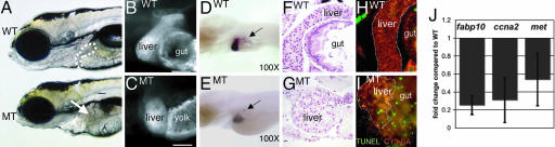
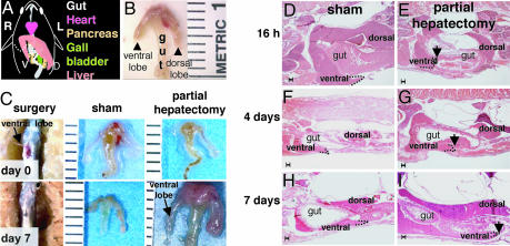
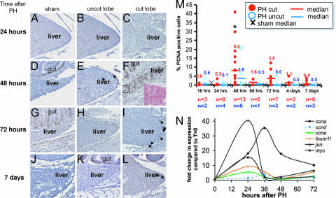
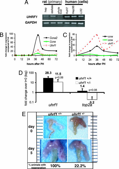
In contrast to the deregulated hepatocellular division that is a feature of many hepatic diseases and malignancies, physiologic liver growth during embryonic development and after partial hepatectomy (PH) in adults is characterized by tightly controlled cell proliferation. We used forward genetic screening in zebrafish to test the hypothesis that a similar genetic program governs physiologic liver growth during hepatogenesis and regeneration after PH. We identified the uhrf1 gene, a cell cycle regulator and transcriptional activator of top2a expression, as required for hepatic outgrowth and embryonic survival. By developing a methodology to perform PH on adult zebrafish, we found that liver regeneration inuhrf1+/- adult animals is impaired.uhrf1 transcript levels dramatically increase after PH in both mice, and zebrafish and top2a is not up-regulated in uhrf1+/- livers after PH. This indicates that uhrf1 is required for physiologic liver growth in both embryos and adults and illustrates that zebrafish livers regenerate.


Conflict of interest statement

The authors declare no conflict of interest.

Figures

Fig. 1.
Fig. 1.
hi272 mutant embryos have a small-for-size liver on day 5. (A) WT and MT liver day 5 embryos from hi272. The liver in phenotypically WT embryo is visible anterior to the intestinal bulb (white outline); the expected position of the liver in the MT is indicated by an arrow. (B and C) CY3-SA labeling of day 5 WT (B) and MT embryos (C). The gut in the mutant is malformed and does not stain with CY3-SA, and the yolk consumption is incomplete in the mutant by day 5, and thus is labeled with CY3-SA. (Scale bar: 50 μm.) (D and E) In situ hybridization with fabp10 and insulin probes on WT (D) and MT (E) embryos. Arrows point to pancreatic islets, labeled with insulin. (F and G) H&E-; stained sagittal sections of livers from WT(F) and MT (G) embryos. Images were taken through the widest section of the left liver lobe. (Scale bar: 10 μm.) (H and I) Apoptotic cells (green) are not seen in the CY3-SA labeled liver (white outline) and gut of WT (H) but are plentiful in MT (I) embryos. (J) Q-PCR on cDNA prepared from day 5 WT and MT embryos from hi272. Expression levels relative to tbp were calculated and shown as fold change compared with phenotypically WT siblings. The experiment was run in triplicate; bars indicate SD.
Fig. 2.
Fig. 2.
uhrf1 is expressed in zones of proliferation during organogenesis and in proliferating adult tissues. (A and B) In situ hybridization at 24 (A) and 34 (B) hours pf shows high levels of uhrf1 expression in rapidly proliferating cells. (CF) At 48 h pf (C), expression has decreased in most tissues except the tectum, retina, and arches, and by 57 h (D), 4 days (E) and 5 days (F) pf, expression is evident in the liver bud and gut (arrowheads). No expression is seen in MT embryos at 4 days pf (E Inset). (G) uhrf1 message was detected in tissues from and adult male zebrafish by Q-PCR by using tbp as a reference.
Fig. 3.
Fig. 3.
Partial hepatectomy results in regeneration within 7 days. (A) Schematic of the gastrointestinal organs, viewed from the animals ventral side. The animal's left (L) and right (R) are labeled as are the dorsal (D) and ventral (V) liver lobes. The dashed line marks the site of resection of the ventral lobe. (B) Dissected gut and liver with a piece of spleen (s) attached. Ruler marks are millimeters. (C) PH in zebrafish is carried out on day 0 (Upper) by creating a small incision through the ventral body wall and the ventral lobe of the liver is resected. By day 7 (Lower), the wound has healed and a ventral lobe is evident in both sham and animals subject to PH. (DI) H&E stained sagittal sections of fish taken at 16 h (D and E) and 4 (F and G) and 7 (H and I) days after PH or sham operation. The tip of the dorsal lobe is outlined with a dotted line and indicated with an arrow in PH samples. (Scale bars: 100 μm.)
Fig. 4.
Fig. 4.
PCNA expression peaks at 48 h after PH. (AL) PCNA immunostaining is indicated by brown nuclei. The gut lies medial to the dorsal liver lobe, serving as a positive control (see D, F, G, and K). Samples taken from sham operated animals (A, D, G, and J), and both the uncut (ventral) lobe (B, E, H, and K) and cut (dorsal) lobe (C, F, I, and L) of animals that underwent PH at 24 h (AC), 48 h (DF), 72 h (GI), and 7 days (JL) after surgery. Arrows indicate PCNA-positive hepatocyte nuclei. (F Inset) A magnification of the boxed region, stained with H&E. (M) The PCNA index was determined from the cut (red circles) and uncut (blue circles) lobes of PH animals, and the median is plotted as a bar with the value indicated above. The median PCNA index for sham animals is illustrated with an ×. ∗, a significant difference in the PCNA index in the cut lobe at 48 h compared with the cut lobe at 24 h (P < 0.0035) and 72 h (P < 0.095). (N) The remaining ventral lobe of zebrafish livers were collected at the indicated times after PH. The expression levels were determined in two fish by using Q-PCR with ef1a as a reference, the values were averaged and are plotted as fold change compared with expression levels in quiescent livers (t = 0) from five fish.
Fig. 5.
Fig. 5.
uhrf1 is required for liver regeneration. (A) uhrf1 message was detected by standard PCR in rat primary hepatocytes, stellate cells, and whole liver tissue, in human hepatocyte cell lines (Huh7 and Hep3B), and in the stellate cell line (LX2) compared with GAPDH. (B and C) Mouse (B) and zebrafish (C) livers were collected at the indicated times after PH. The expression levels of uhrf1, ccne, and ccna2 were determined by using Q-PCR with cyclophilin as a reference for mouse and ef1a as a reference for zebrafish. Graphs represent fold change compared with quiescent livers. For zebrafish, n = 2 for each PH time point and n = 5 for quiescent liver samples. (D) The level of uhrf1 and top2a transcripts in quiescent (t = 0; n = 3) and regenerating (t = 36 h; n = 3) livers were determined by Q-PCR with ef1a as a reference. Numbers are expressed as the average fold change at 36 h compared with the t = 0 samples. Error bars indicate SD, and P values are labeled. (E) Seven uhrf1+/+ fish and nine uhrf1+/− fish were subject to PH. After 5 days of recovery, the livers were dissected and assessed for regrowth. Representative animals are shown. One hundred percent (7/7) of the uhrf1+/+ animals had substantial regeneration, whereas only 22% (2/7) of the uhrf1+/− animals had any regrowth 5 days after PH.

References

    1. Bucher NL, Swaffield MN. Cancer Res. 1964;24:1611–1625. - PubMed
    1. Taub R. Nat Rev Mol Cell Biol. 2004;5:836–847. - PubMed
    1. Behrens A, Sibilia M, David JP, Mohle-Steinlein U, Tronche F, Schutz G, Wagner EF. EMBO J. 2002;21:1782–1790. - PMC - PubMed
    1. Hilberg F, Aguzzi A, Howells N, Wagner EF. Nature. 1993;365:179–181. - PubMed
    1. Johnson RS, van Lingen B, Papaioannou VE, Spiegelman BM. Genes Dev. 1993;7:1309–1317. - PubMed

Publication types

MeSH terms