. 2007 Mar;63(5):1331-44.
doi: 10.1111/j.1365-2958.2007.05592.x. Epub 2007 Jan 22.

Scavenging of the cofactor lipoate is essential for the survival of the malaria parasite Plasmodium falciparum

Affiliations

Scavenging of the cofactor lipoate is essential for the survival of the malaria parasite Plasmodium falciparum

Marina Allary et al. Mol Microbiol. 2007 Mar.

Abstract

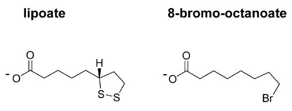
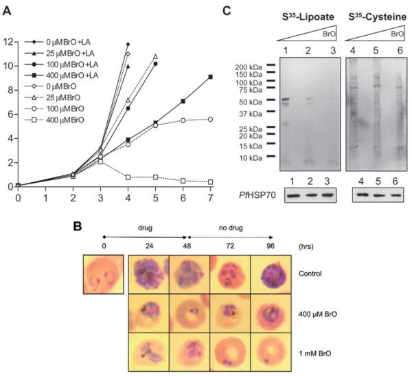
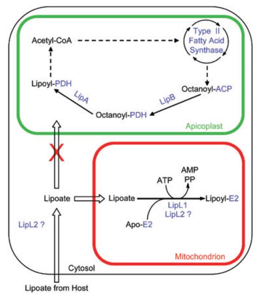
Lipoate is an essential cofactor for key enzymes of oxidative metabolism. Plasmodium falciparum possesses genes for lipoate biosynthesis and scavenging, but it is not known if these pathways are functional, nor what their relative contribution to the survival of intraerythrocytic parasites might be. We detected in parasite extracts four lipoylated proteins, one of which cross-reacted with antibodies against the E2 subunit of apicoplast-localized pyruvate dehydrogenase (PDH). Two highly divergent parasite lipoate ligase A homologues (LplA), LipL1 (previously identified as LplA) and LipL2, restored lipoate scavenging in lipoylation-deficient bacteria, indicating that Plasmodium has functional lipoate-scavenging enzymes. Accordingly, intraerythrocytic parasites scavenged radiolabelled lipoate and incorporated it into three proteins likely to be mitochondrial. Scavenged lipoate was not attached to the PDH E2 subunit, implying that lipoate scavenging drives mitochondrial lipoylation, while apicoplast lipoylation relies on biosynthesis. The lipoate analogue 8-bromo-octanoate inhibited LipL1 activity and arrested P. falciparum in vitro growth, decreasing the incorporation of radiolabelled lipoate into parasite proteins. Furthermore, growth inhibition was prevented by lipoate addition in the medium. These results are consistent with 8-bromo-octanoate specifically interfering with lipoate scavenging. Our study suggests that lipoate metabolic pathways are not redundant, and that lipoate scavenging is critical for Plasmodium intraerythrocytic survival.


Figures

Fig. 1
Fig. 1
The structures of R-lipoate and BrO.
Fig. 2
Fig. 2
Evidence of lipoylation and lipoate scavenging in P. falciparum. A. Western blot analysis of a lysate from asynchronous erythrocytic stage parasites probed with antiserum specific for lipoylated proteins (1:10 000). B. Western blot analysis of a lysate from asynchronous erythrocytic stage parasites probed with antibodies specific for the E2 subunit of P. falciparum PDH (1:500). C. Incorporation of radiolabelled lipoate into proteins. Erythrocytic stage parasites were cultured for 2 days in the presence of [35S]-lipoate (0.3 μCi ml−1). Protein extracts were separated by SDS-PAGE and analysed by autoradiography. The putative assignment of protein bands is indicated.
Fig. 3
Fig. 3
P. falciparum possesses two functional LplA homologues. A. CLUSTALW alignment of P. falciparum LipL1 and LipL2 amino acid sequences. Boxed residues correspond to the conserved lipoate ligase region (KOG3159). The amino acids involved in the interaction with the lipoyl-AMP intermediate in the crystal structure of Thermoplasma acidophilum LplA (Kim et al., 2005) are highlighted in bold. Triangles mark the first amino acids in the LipL120 and LipL229 constructs. B. Functional complementation of lipoylation-deficient E. coli strain TM136. TM136 cells transformed with mature LipL1 (L120), full-length LipL2 (L2FL), a putative mature LipL2 (L229), or the pMAL vector alone were incubated at 37°C in non-permissive minimal E medium containing 0.1 mM IPTG, in the presence or in the absence of 10 ng ml−1 lipoate. The starting optical density (OD600) for each culture was 0.01. Cell growth was assessed by measuring the OD600 of the cultures after 48 h (open bars) and 72 h (closed bars). Experiments were conducted in triplicate and error bars indicate the standard deviation. C. LipL1 and LipL2 specificity for E. coli lipoate acceptor proteins. After functional complementation, lysates from equivalent amounts of cells expressing LipL120, LipL2FL and LipL229 were analysed by Western blot using antiserum specific for lipoylated proteins (α-LA). The absence (−) or presence (LA) of lipoate in the complementation medium is indicated. Loading was normalized to the optical density. The lipoylation pattern of the BL21 strain, which is wild-type for lipoylation (WT, about five times less cells), and of the TM136 cells expressing the pMAL vector alone [pMAL, requiring succinate and acetate (SA) for growth] are also shown. The same blot was stripped and reprobed with antiserum specific for E. coli DnaK as a loading control (α-DnaK).
Fig. 4
Fig. 4
Inhibition by BrO of pure recombinant LipL1 enzymatic activity. A. Coomassie-stained SDS-PAGE gel showing purified LipL120. B. LipL1 lipoylation activity. Purified LipL120 (0.3 μM) was assayed for lipoate ligase activity in the presence (+) or the absence of several reaction components as indicated, using 1.8 μM apo-PfH-protein (upper panel) or 2 μM apo-EcH-protein (lower panel). The lipoate concentration was 180 μM. The reactions were analysed by SDS-PAGE, and lipoylated proteins were detected by Western blot analysis using antiserum specific for lipoylated proteins. X, heat-killed LipL120. C. LipL1 octanoylation activity. Purified LipL120 (3 μM) was assayed for octanoate ligase activity in the presence (+) or the absence (−) of 1.8 mM ATP using 10 μM apo-PfH-protein (upper panel) or apo-EcH-protein (lower panel). The [1-14C]-octanoate concentration was 72 μM. The reactions were analysed by SDS-PAGE, and octanoylated proteins were detected by autoradiography. D. Inhibition of LipL1 lipoylation activity by BrO. LipL120 was assayed as described in B with apo-PfH-protein as substrate in the presence of 180 μM lipoate and 0, 1 and 10 mM BrO. The percentage of inhibition as compared with the control reaction without BrO is indicated below the figure. E. Inhibition of LipL1 octanoylation activity by BrO. LipL120 was assayed as described in C with apo-PfH-protein as substrate in the presence of 72 μM [1-14C]-octanoate and of various concentrations of BrO. Octanoylated H-protein was TCA-precipitated and quantified by scintillation counting. The graph shows the percentage inhibition versus BrO concentration.
Fig. 5
Fig. 5
Effects of BrO on P. falciparum cultures. A. Effect of BrO on P. falciparum proliferation. Asynchronous parasites were cultured for 7 days in the presence of 0, 25, 100 and 400 μM BrO, with or without additional 2 μM lipoate (LA) in the growth medium. Cultures were monitored and their parasitaemia assessed with daily Giemsa-stained smears. B. Effect of BrO on synchronized parasites. P. falciparum cultures at the ring stage were incubated with 400 μM and 1 mM BrO for 48 h with daily medium change, then subsequently maintained without the lipoate analogue. Images of Giemsa-stained parasites from the different cultures analysed every 24 h are shown. C. Effect of BrO on parasite incorporation of exogenous lipoate. Asynchronous cultures were grown for 2 days in the absence (lanes 1 and 4) or in the presence of 100 μM (lanes 2 and 5) and 400 μM (lanes 3 and 6) BrO. The culture media also contained [35S]-lipoate (0.9 μCi ml−1) or as a control, [35S]-cysteine (20 μCi ml−1), as indicated. Parasite extracts were analysed by SDS-PAGE followed by autoradiography (upper panels) or by Western blot using anti-PfHSP70 antibodies (lower panels).
Fig. 6
Fig. 6
Model of lipoate metabolism in P. falciparum erythrocytic parasites. The scheme describes lipoate synthesis in the apicoplast and lipoate scavenging in the mitochondrion. Proteins are coloured blue. In the mitochondrion, ‘E2′ could represent the H-protein or the E2 subunits of BCDH or KGDH. The two putative locations of LipL2 are shown.

References

    1. Bahl A, Brunk B, Crabtree J, Fraunholz MJ, Gajria B, Grant GR, et al. PlasmoDB: the Plasmodium genome resource. A database integrating experimental and computational data. Nucleic Acids Res. 2003;31:212–215. - PMC - PubMed
    1. Bender A, van Dooren GG, Ralph SA, McFadden GI, Schneider G. Properties and prediction of mitochondrial transit peptides from Plasmodium falciparum. Mol Biochem Parasitol. 2003;132:59–66. - PubMed
    1. Booker SJ. Unraveling the pathway of lipoic acid biosynthesis. Chem Biol. 2004;11:10–12. - PubMed
    1. Bryk R, Lima CD, Erdjument-Bromage H, Tempst P, Nathan C. Metabolic enzymes of mycobacteria linked to antioxidant defense by a thioredoxin-like protein. Science. 2002;295:1073–1077. - PubMed
    1. Bunik VI. 2-Oxo acid dehydrogenase complexes in redox regulation. Eur J Biochem. 2003;270:1036–1042. - PubMed

Publication types

MeSH terms

Associated data

LinkOut - more resources