

Participation of the Fas and Fas ligand systems in apoptosis during atrophy of the rat submandibular glands
- PMID: 17244334
- PMCID: PMC2517292
- DOI: 10.1111/j.1365-2613.2006.00511.x
Participation of the Fas and Fas ligand systems in apoptosis during atrophy of the rat submandibular glands
Abstract
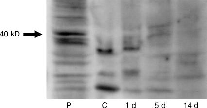
Most acinar cells and some duct cells undergo apoptosis during atrophy of the submandibular gland. The present study was designed to elucidate whether Fas and its receptor ligand (FasL) are involved during apoptotic atrophy of the gland. The excretory duct of the right submandibular gland of rats was doubly ligated with metal clips from 1 to 14 days for induction of gland atrophy. Control rats were untreated. Fas and FasL expression in the atrophied submandibular gland was detected using immunohistochemistry (IHC) and Western immunoblot. Expression of activated caspase 8 and activated caspase 3 was also detected with IHC. Fas-positive acinar and duct cells and FasL-positive duct cells increased in the atrophic glands at 3 and 5 days after duct ligation when apoptotic cells were commonly observed. Thereafter, Fas- and FasL-positive cells declined in number. Patterns of expression of Fas and FasL using Western immunoblots concurred with the IHC results. Activated caspase 8-positive cells were present at every time interval but peaked at 3 and 5 days following duct ligation. The cells showing immunoreaction for activated caspase 3 first appeared on day 3, with the peak in apoptosis, after which they decreased. The results indicate that the Fas/FasL systems likely play an important role in apoptotic pathways during atrophy of the submandibular gland.
Figures


Similar articles
-
Cellular expression of Bcl-2 and Bax in atrophic submandibular glands of rats.Int J Exp Pathol. 2008 Oct;89(5):303-8. doi: 10.1111/j.1365-2613.2008.00613.x. Int J Exp Pathol. 2008. PMID: 18808524 Free PMC article.
-
Mitotic proliferation of myoepithelial cells during regeneration of atrophied rat submandibular glands after duct ligation.J Oral Pathol Med. 2004 Aug;33(7):430-4. doi: 10.1111/j.1600-0714.2004.00234.x. J Oral Pathol Med. 2004. PMID: 15250836
-
Apoptosis and proliferation of myoepithelial cells in atrophic rat submandibular glands.J Histochem Cytochem. 2001 Dec;49(12):1557-64. doi: 10.1177/002215540104901209. J Histochem Cytochem. 2001. PMID: 11724903
-
Cell death and cell proliferation in the regeneration of atrophied rat submandibular glands after duct ligation.J Oral Pathol Med. 2004 Jan;33(1):23-9. doi: 10.1111/j.1600-0714.2004.00191.x. J Oral Pathol Med. 2004. PMID: 14675137
-
Mitochondrial- and Fas-L-mediated pathways involved in quinestrol induced spermatogenic apoptosis in adult rat testes.Toxicol Mech Methods. 2014 Dec;24(9):609-15. doi: 10.3109/15376516.2014.970680. Epub 2014 Oct 10. Toxicol Mech Methods. 2014. PMID: 25258304 Review.
Cited by
-
Regeneration of acinar cells following ligation of rat submandibular gland retraces the embryonic-perinatal pathway of cytodifferentiation.Differentiation. 2010 Feb;79(2):120-30. doi: 10.1016/j.diff.2009.11.005. Epub 2010 Jan 6. Differentiation. 2010. PMID: 20056310 Free PMC article.
-
P2 Receptors as Therapeutic Targets in the Salivary Gland: From Physiology to Dysfunction.Front Pharmacol. 2020 Mar 13;11:222. doi: 10.3389/fphar.2020.00222. eCollection 2020. Front Pharmacol. 2020. PMID: 32231563 Free PMC article. Review.
-
Cellular expression of Bcl-2 and Bax in atrophic submandibular glands of rats.Int J Exp Pathol. 2008 Oct;89(5):303-8. doi: 10.1111/j.1365-2613.2008.00613.x. Int J Exp Pathol. 2008. PMID: 18808524 Free PMC article.
-
Duct ligation/de-ligation model: exploring mechanisms for salivary gland injury and regeneration.Front Cell Dev Biol. 2024 Jun 25;12:1399934. doi: 10.3389/fcell.2024.1399934. eCollection 2024. Front Cell Dev Biol. 2024. PMID: 38983787 Free PMC article. Review.
-
Experimental Animal Model Systems for Understanding Salivary Secretory Disorders.Int J Mol Sci. 2020 Nov 10;21(22):8423. doi: 10.3390/ijms21228423. Int J Mol Sci. 2020. PMID: 33182571 Free PMC article. Review.
References
-
- Barnhart BC, Alappat EC, Peter ME. The CD95 Type I/Type II model. Semin. Immunol. 2003;15:185–193. - PubMed
-
- Bellgrau D, Gold D, Selawry H, Moore J, Franzusoff A, Duke RC. A role for CD95 ligand in preventing graft rejection. Nature. 1995;377:630–632. - PubMed
-
- Cummins M, Dardick I, Burford-Mason A. Obstructive sialadenitis: a rat model. J. Otolaryngol. 1994;23:50–56. - PubMed
-
- Curtin JF, Cotter TG. Live and let die: regulatory mechanisms in Fas-mediated apoptosis. Cell Signal. 2003;15:983–992. - PubMed
Publication types
MeSH terms
Substances
LinkOut - more resources
Full Text Sources
Research Materials
Miscellaneous
