. 2007 Jan 26;25(2):207-17.
doi: 10.1016/j.molcel.2007.01.001.

RACK1 competes with HSP90 for binding to HIF-1alpha and is required for O(2)-independent and HSP90 inhibitor-induced degradation of HIF-1alpha

Affiliations

RACK1 competes with HSP90 for binding to HIF-1alpha and is required for O(2)-independent and HSP90 inhibitor-induced degradation of HIF-1alpha

Ye V Liu et al. Mol Cell. .

Abstract

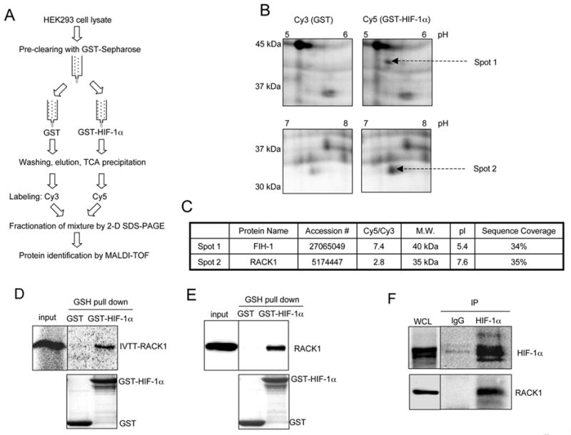
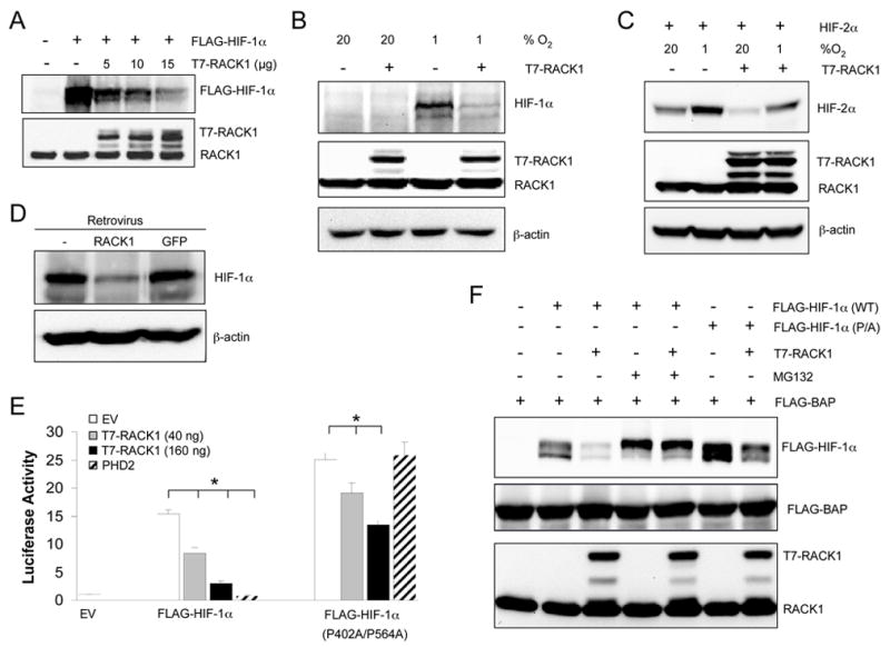
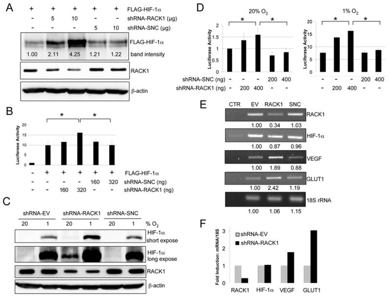
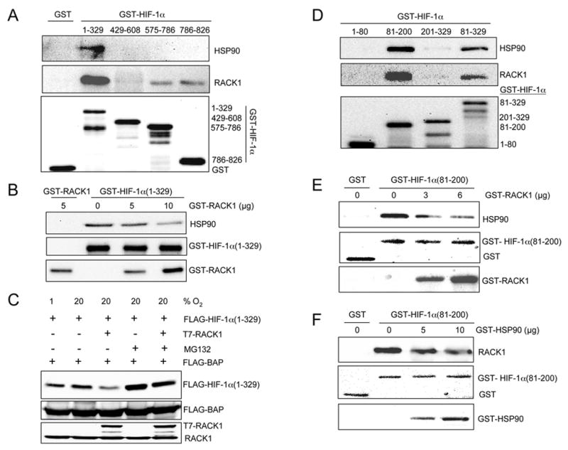
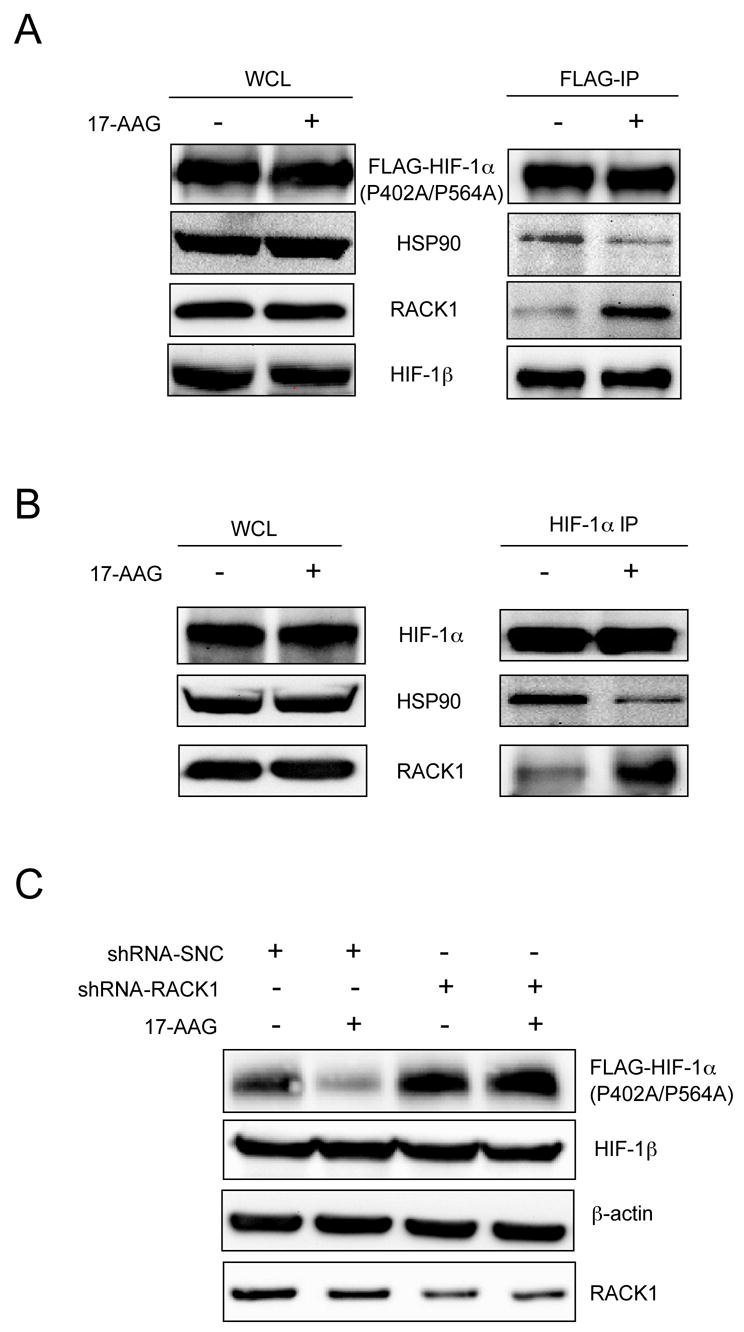
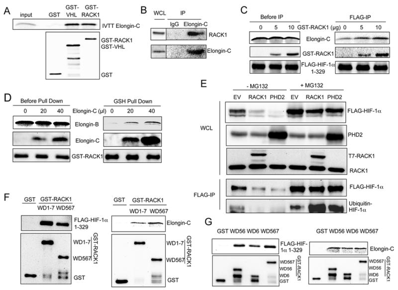
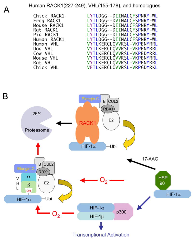
Hypoxia-inducible factor 1 (HIF-1) regulates transcription in response to changes in O(2) concentration. O(2)-dependent degradation of the HIF-1alpha subunit is mediated by prolyl hydroxylase (PHD), the von Hippel-Lindau (VHL)/Elongin-C/Elongin-B E3 ubiquitin ligase complex, and the proteasome. Inhibition of heat-shock protein 90 (HSP90) leads to O(2)/PHD/VHL-independent degradation of HIF-1alpha. We have identified the receptor of activated protein kinase C (RACK1) as a HIF-1alpha-interacting protein that promotes PHD/VHL-independent proteasomal degradation of HIF-1alpha. RACK1 competes with HSP90 for binding to the PAS-A domain of HIF-1alpha in vitro and in human cells. HIF-1alpha degradation induced by the HSP90 inhibitor 17-allylaminogeldanamycin is abolished by RACK1 loss of function. RACK1 binds to Elongin-C and promotes ubiquitination of HIF-1alpha. Elongin-C-binding sites in RACK1 and VHL show significant sequence similarity. Thus, RACK1 is an essential component of an O(2)/PHD/VHL-independent mechanism for regulating HIF-1alpha stability through competition with HSP90 and recruitment of the Elongin-C/B ubiquitin ligase complex.


Figures

Figure 1
Figure 1. Identification of RACK1 as a HIF-1α Interacting Protein
(A) Proteomics strategy employed to identify proteins that interact specifically with a GST fusion protein containing residues 531–826 of human HIF-1α. MALDI-TOF, matrix assisted laser desorption ionization-time-of-flight mass spectrometry. (B) Fluorescence images of spot 1 and spot 2 from the 2-D gel. (C) Protein identification for spot 1 and spot 2. Accession #, NCBI Protein Database. Cy5/Cy3, ratio of fluorescence intensities. Sequence Coverage, MALDI-TOF data. (D) Binding assay using in vitro translated RACK1. Purified GST or GST-HIF-1α (531–826) was: incubated with in vitro transcribed, translated, and 35S-labeled RACK1 (IVTT- RACK1); captured on glutathione (GSH)-Sepharose beads; and analyzed by SDS-PAGE and autoradiography (top panel). An aliquot of RACK1 was also applied directly to the gel (input). The GST fusion proteins were visualized on the gel by Coomassie blue staining (bottom panel) (E) Binding assay using cell lysates. Purified GST or GST-HIF-1α (531–826) was incubated with HEK293 lysate, captured on GSH-Sepharose beads, and analyzed by immunoblot (IB) assay using antibody (Ab) against RACK1 (top panel). An aliquot of cell lysate was also applied directly to the gel (input). The GST fusion proteins were visualized on the blot by Ponceau S staining (bottom panel). (F) Co-immunoprecipitation (co-IP) of endogenous RACK1 and HIF-1α. IP was performed using whole cell lysates (WCL), which were prepared from HEK293 cells treated for 4 hr with 100 μM desferrioxamine, and either mouse IgG or anti-HIF-1α Ab. WCL and IP products were subjected to IB assays using Ab against HIF-1α or RACK1.
Figure 2
Figure 2. RACK1 Inhibits HIF-1α Protein Expression by a Mechanism that is Independent of Prolyl Hydroxylation but Dependent on Proteasome Activity
(A) RACK1 reduces HIF-1α protein levels in cotransfected cells. HEK293T cells were cotransfected with empty vector (EV) or expression vector encoding FLAG-HIF-1α or T7-RACK1. WCL were subjected to IB assay to detect FLAG-HIF-1α, T7-RACK1, and endogenous RACK1. (B) RACK1 reduces levels of endogenous HIF-1α protein in hypoxic cells. HEK293T cells were transfected with T7-RACK1 or EV. The cells were exposed to 20% or 1% O2 for 4 hr. WCL were subjected to IB assay to detect T7-RACK1 and endogenous HIF-1α, RACK1, and β-actin. (C) RACK1 reduces HIF-2α protein levels. HEK293T cells were cotransfected with vector encoding HIF-2α and T7-RACK1 or EV. The cells were exposed to 20% or 1% O2 for 4 hr. WCL were subjected to IB assay to detect HIF-2α, RACK1, and β-actin. (D) RACK1 reduces endogenous HIF-1α protein levels in VHL-deficient renal carcinoma cells. RCC4 cells were transduced with retrovirus encoding RACK1 or GFP. WCL from parental RCC4 cells and retrovirus-infected cells were subjected to IB assay to detect endogenous HIF-1α and β-actin. (E) RACK1, but not PHD2, inhibits transcription mediated by hydroxylation-resistant HIF-1α. HEK293T cells were cotransfected with: pSV-Renilla, in which Renilla luciferase is expressed from an SV40 promoter; p2.1, which contains a hypoxia response 31 element (HRE) upstream of an SV40 promoter and firefly luciferase coding sequences; EV or expression vectors encoding wild-type FLAG-HIF-1α or FLAG-HIF-1α(P402A/P564A); and T7-RACK1 or PHD2 expression vector. After 24 hr, cells were lysed and the ratio of firefly:Renilla luciferase activity was determined. The results were normalized to those from cells transfected with EV (mean + SE shown). * P < 0.01 compared to EV. (F) RACK1 induces proteasomal degradation of wild-type or hydroxylation-deficient HIF-1α. Cells were cotransfected with wild-type (WT) or proline-to-alanine substituted (P/A) FLAG-HIF-1α vector, T7-RACK1 or EV, and FLAG-tagged bacterial alkaline phosphatase (FLAG-BAP) vector. Transfected cells were treated with vehicle or MG132 (10 μM) for 4 hr. WCL were subjected to IB assay to detect FLAG-HIF-1α, FLAG-BAP, and RACK1.
Figure 3
Figure 3. RACK1 Knockdown by RNA Interference Increases HIF-1α Protein Levels and HIF-1 Transcriptional Activity
(A) Effect of RACK1 knockdown on FLAG-HIF-1α levels. HEK293T cells were cotransfected with FLAG-HIF-1α and short hairpin RNA (shRNA) against RACK1 or a scrambled negative control (SNC). WCL were collected 48 hr after transfection and analyzed by IB assays to detect FLAG-HIF-1α, endogenous RACK1, and β-actin. FLAG-HIF-1α levels were quantified by densitometric analysis (band intensity). (B) HEK293T cells were cotransfected with: pSV-Renilla; p2.1; FLAG-HIF-1α; and shRNA-SNC, shRNA-RACK1, or EV. After 24 hr, cells were lysed and the ratio of firefly:Renilla luciferase activity was determined. The results were normalized to those from cells transfected with EV (mean + SE shown). * P < 0.01 for indicated comparisons. (C) Effect of RACK1 knockdown on endogenous HIF-1α levels. Cells were transfected with shRNA-RACK1, shRNA-SNC, or EV. After 68 hours, cells were exposed to 20% or 1% O2 for 4 hr. WCL were collected and analyzed by IB assays to detect endogenous HIF-1α, RACK1, and β-actin. Longer exposure of the HIF-1α blot revealed HIF-1α levels at 20% O2. (D) HEK293T cells were cotransfected with: pSV-Renilla; p2.1; shRNA-SNC, shRNA-RACK1, or EV. After 24 hr, cells were exposed to 20% or 1% O2 for another 24 hr and the ratio of firefly:Renilla luciferase activity was determined (mean + SE shown). *P < 0.05 for indicated comparisons. (E) Effect of RACK1 knockdown on mRNA expression. Cells were transfected with EV, shRNA-SNC, or shRNA-RACK1 expression vector. 48 hr after transfection, RNA was isolated and RT-PCR was performed to amplify RACK1, HIF-1α, VEGF, GLUT1 mRNA and 18S rRNA. CTR, control in which reverse transcriptase was omitted from the cDNA synthesis reaction. Band intensities were quantified by densitometry relative to EV. (F) Cells were transfected with EV or shRNA-RACK1 expression vector. 48 hr after transfection, RNA was isolated and real-time RT-PCR was performed. The ratio of mRNA to 18S rRNA was normalized to EV for each mRNA (mean + SE shown).
Figure 4
Figure 4. RACK1 Competes with HSP90 for Binding to HIF-1α
(A) Binding of RACK1 and HSP90 to HIF-1α. GST or GST-HIF-1α fusion protein (encoding HIF-1α residues 1–329, 429–608, 575–786, or 786–826) was incubated with HEK293T cell lysate. The GST fusion proteins were captured by GSH-Sepharose beads, and analyzed by IB assays to detect bound HSP90, RACK1, and GST-HIF-1α. (B) RACK1 and HSP90 compete for binding to HIF-1α (1–329). GST-RACK1 or GST-HIF-1α(1–329) was tested for binding to HSP90 present in WCL. In lanes 2–4, 0, 5 or 10 μg of GST-RACK1 was added to compete with HSP90 for binding to GST-HIF-1α. GST fusion proteins were captured by GSH-Sepharose beads, and analyzed by IB assay to detect bound HSP90, GST-HIF-1α, and GST-RACK1. (C) RACK1 induces proteasomal degradation of HIF-1α (1–329). HEK293T cells were cotransfected with FLAG-BAP, FLAG-HIF-1α (1–329), and T7-RACK1 or EV. Transfected cells were exposed to 20% or 1% O2 in the presence of vehicle or MG132 (10 μM) for 4 hr. IB assays were performed to detect FLAG-HIF-1α(1–329), FLAG-BAP, and RACK1 in WCL. (D) HIF-1α residues 81–200 are sufficient for binding to RACK1 or HSP90. GST-HIF-1α fusion protein was incubated with WCL, captured by GSH-Sepharose beads, and analyzed by IB assays to detect bound HSP90, RACK1, and GST-HIF-1α. (E) RACK1 competes with HSP90 for binding to HIF-1α (81–200). 3 μg of GST or GST-HIF-1α (81–200) was incubated with HEK293T lysate. 0, 3 or 6 μg of GST-RACK1 was added to compete with HSP90 for binding to GST-HIF-1α. GST fusion proteins were captured by GSH-Sepharose beads, and analyzed by IB assay to detect bound HSP90. GST proteins were visualized on the blot by Ponceau S staining. (F) HSP90 competes with RACK1 for binding to HIF-1α (81–200). 3 μg of GST or GST-HIF-1α(81–200) was incubated with WCL. 0, 5 or 10 μg of GST-HSP90 was added to compete with RACK1 for binding to GST-HIF-1α. GST fusion proteins were captured by GSH-Sepharose beads and analyzed by IB assay to detect bound RACK1. GST proteins were visualized on the blot by Ponceau S staining.
Figure 5
Figure 5. HIF-1α Degradation Induced by 17-AAG is Dependent on RACK1 Binding
(A) 17-AAG treatment decreases HSP90 binding and increases RACK1 binding to HIF-1α in cells. HEK293T cells were transfected with FLAG-HIF-1α (P402A/P564A). 24 hr after transfection, 0.5 μM 17-AAG or vehicle was added to the cells. 40 hr after transfection, 5 μM MG132 was added to the cells for 8 hr. WCL were subject to IP with anti-FLAG. The WCL and IP product were subjected to IB assay to detect FLAG-HIF-1α, HSP90, RACK1, and HIF-1β. (B) 17-AAG treatment decreases HSP90 binding and increases RACK1 binding to HIF-1α in vitro. Cells were exposed to 1% O2 for 4 hr. WCL were treated with vehicle or 2 μM 17-AAG for 2 hr at 4 °C followed by IP with anti-HIF-1α Ab. The WCL and IP products were subjected to IB assay to detect HIF-1α, HSP90, and RACK1. (C) RACK1 knockdown blocks HIF-1α degradation induced by 17-AAG. Cells were cotransfected with FLAG-HIF-1α (P402A/P564A) and shRNA-RACK1 or shRNA-SNC. 24 hr later, 0.5 μM 17-AAG or vehicle was added. After 24 hr, cells were harvested and IB assays were performed to detect FLAG-HIF-1α, HIF-1β, β-actin, and RACK1.
Figure 6
Figure 6. RACK1 Promotes Elongin-C Binding to HIF-1α and Ubiquitination of HIF-1α
(A) Purified GST proteins were incubated with 35S-labeled Elongin-C, captured on GSH-Sepharose beads, and fractionated by SDS-PAGE. Bound Elongin-C and GST fusion proteins were analyzed by autoradiography and anti-GST IB respectively. (B) WCL were subjected to IP with goat IgG or anti-Elongin-C Ab. The WCL and IP products were analyzed by IB to detect endogenous Elongin-C and RACK1. (C) WCL from FLAG-HIF-1α(1–329)-transfected cells was incubated with 35S-Elongin-C and 0, 5, or 10 μg of GST-RACK1, followed by anti-FLAG IP. The mixture before IP (left panel) and IP product (right panel) were analyzed by SDS-PAGE followed by autoradiography (Elongin-C) and IB (GST-RACK1 and FLAG-HIF-1α). (D) 10 μl of 35S-Elongin-B was incubated with: GST-RACK1; 0, 20 or 40 μl of unlabelled Elongin-C; and 40, 20 or 0 μl of unprogrammed reticulate lysate. GST-RACK1 was captured by GSH-Sepharose beads. The mixtures before (left panel) and after (right panel) GSH capture were analyzed by SDS-PAGE followed by autoradiography (Elongin-B) and IB (GST-RACK1 and Elongin-C). (E) HEK293T cells were transfected with FLAG-HIF-1α and T7-RACK1, PHD2, or EV. 24 hr after transfection, cells were treated with vehicle or 10 μM MG132 for 4 hr. WCL were collected and IP was performed with anti-FLAG Ab. IB assays were used to detect FLAG-HIF-1α, PHD2, and RACK1. An anti-ubiquitin Ab was used to detect ubiquitinated HIF-1α after FLAG IP. (F)Purified GST and GST-RACK1 (WD1-7 or WD5-7) was incubated with WCL from FLAG-HIF-1α (1–329)-transfected HEK293T cells or with 35S-Elongin-C. GST fusion proteins were captured by GSH-Sepharose beads. Bound FLAG-HIF-1α and Elongin-C were detected by anti-FLAG IB (top left panel) was autoradiography (top right panel). Bottom panels, anti-GST IB. (G) GST proteins were incubated with WCL from FLAG-HIF-1α(1–329)-transfected cells (left panels) or with 35S-Elongin-C (right panels) and captured by GSH-Sepharose beads. Bound FLAG-HIF-1α and Elongin-C were detected by anti-FLAG IB (top left panel) was autoradiography (top right panel). Bottom panels, anti-GST IB.
Figure 7
Figure 7. Structural and Functional Similarities Between RACK1 and VHL
(A) Aligned amino acid sequences from RACK1 WD6 domain and VHL α domain. Identical (blue), conserved hydrophobic (green) and conserved aromatic (purple) residues are highlighted. (B) Both an O2-dependent pathway, involving VHL, and an O2-independent pathway, involving RACK1 competition with HSP90, lead to Elongin-C/B recruitment, ubiquitination, and proteasomal degradation of HIF-1α.

Similar articles

Cited by

References

    1. Baek JH, Mahon PC, Oh J, Kelly B, Krishnamachary B, Pearson M, Chan DA, Giaccia AJ, Semenza GL. OS-9 interacts with hypoxia-inducible factor 1α and prolyl hydroxylases to promote oxygen-dependent degradation of HIF-1α. Mol Cell. 2005;17:503–512. - PubMed
    1. Berra E, Benizri E, Ginouves A, Volmat V, Roux D, Pouyssegur J. HIF prolyl-hydroxylase 2 is the key oxygen sensor setting low steady-state levels of HIF-1α in normoxia. EMBO J. 2003;22:4082–4090. - PMC - PubMed
    1. Bruick RK, McKnight SL. A conserved family of prolyl-4-hydroxylases that modify HIF. Science. 2001;294:1337–1340. - PubMed
    1. Ceci M, Gaviraghi C, Gorrini C, Sala LA, Offenhauser N, Marchisio PC, Biffo S. Release of eIF6 (p27BBP) from the 60S subunit allows 80S ribosome assembly. Nature. 2003;426:579–584. - PubMed
    1. Elvidge GP, Glenny L, Appelhoff RJ, Ratcliffe PJ, Ragoussis J, Gleadle JM. Concordant regulation of gene expression by hypoxia and 2-oxoglutarate-dependent dioxygenase inhibition: the role of HIF-1α, HIF-2α, and other pathways. J Biol Chem. 2006;281:15215–15226. - PubMed

Publication types

MeSH terms

LinkOut - more resources