. 2007 Mar 23;282(12):8724-33.
doi: 10.1074/jbc.M609613200. Epub 2007 Jan 23.

Interleukin-1-induced NF-kappaB activation is NEMO-dependent but does not require IKKbeta

Affiliations

Interleukin-1-induced NF-kappaB activation is NEMO-dependent but does not require IKKbeta

Laura A Solt et al. J Biol Chem. .

Abstract

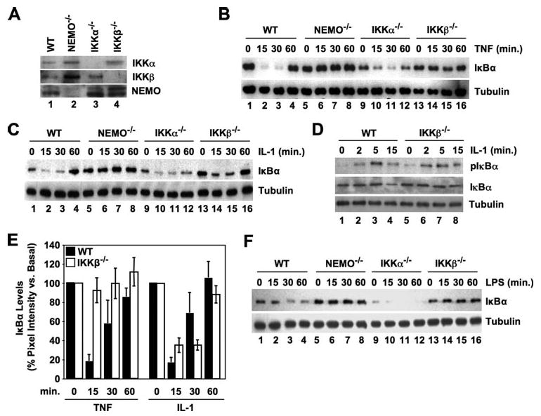
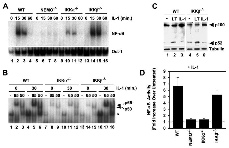
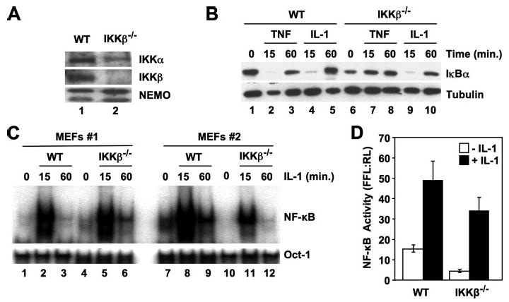
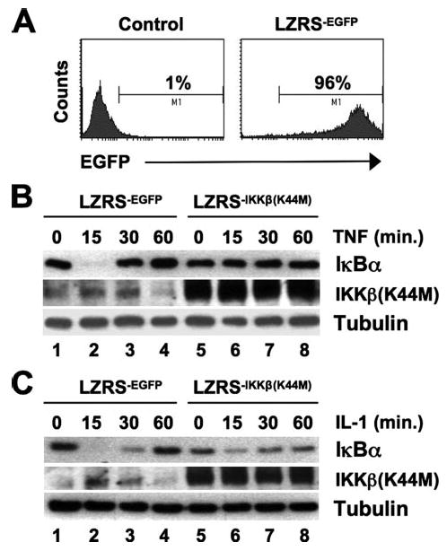
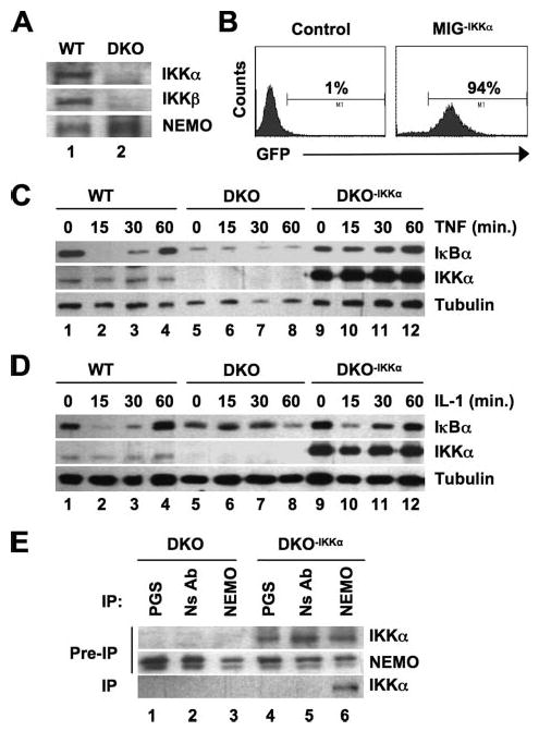
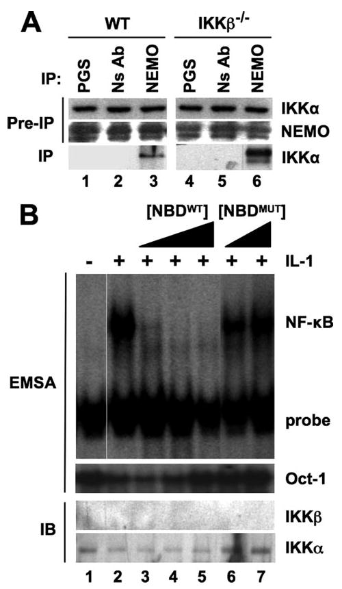
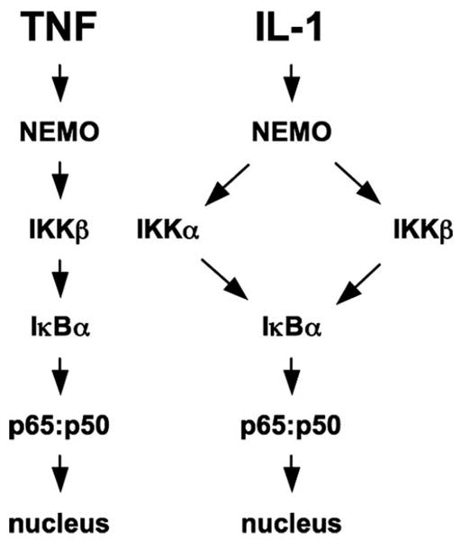
Activation of NF-kappaB by the pro-inflammatory cytokines tumor necrosis factor (TNF) and interleukin-1 (IL-1) requires the IkappaB kinase (IKK) complex, which contains two kinases named IKKalpha and IKKbeta and a critical regulatory subunit named NEMO. Although we have previously demonstrated that NEMO associates with both IKKs, genetic studies reveal that only its interaction with IKKbeta is required for TNF-induced NF-kappaB activation. To determine whether NEMO and IKKalpha can form a functional IKK complex capable of activating the classical NF-kappaB pathway in the absence of IKKbeta, we utilized a panel of mouse embryonic fibroblasts (MEFs) lacking each of the IKK complex subunits. This confirmed that TNF-induced IkappaBalpha degradation absolutely requires NEMO and IKKbeta. In contrast, we consistently observed intact IkappaBalpha degradation and NF-kappaB activation in response to IL-1 in two separate cell lines lacking IKKbeta. Furthermore, exogenously expressed, catalytically inactive IKKbeta blocked TNF- but not IL-1-induced IkappaBalpha degradation in wild-type MEFs, and reconstitution of IKKalpha/beta double knockout cells with IKKalpha rescued IL-1- but not TNF-induced NF-kappaB activation. Finally, we have shown that incubation of IKKbeta-deficient MEFs with a cell-permeable peptide that blocks the interaction of NEMO with the IKKs inhibits IL-1-induced NF-kappaB activation. Our results therefore demonstrate that NEMO and IKKalpha can form a functional IKK complex that activates the classical NF-kappaB pathway in response to IL-1 but not TNF. These findings further suggest NEMO differentially regulates the fidelity of the IKK subunits activated by distinct upstream signaling pathways.


Figures

FIGURE 1
FIGURE 1. IL-1 (but not TNF or LPS) induces IκBα degradation in IKKβ−/− MEFs
A, lysates from wild-type (WT), NEMO-deficient, IKKα−/−, and IKKβ−/− MEFs were analyzed by immunoblotting using the antibodies indicated. B, C, and F, the MEFs indicated (top) were either untreated or incubated with TNF (10 ng/ml) (B), IL-1 (10 ng/ml) (C), or LPS (100 ng/ml) (F) for the times shown, and then lysates were prepared and immunoblotted using anti-IκBα (upper panels). The same blots were also probed using anti-tubulin as a loading control (lower panels). D, WT and IKKβ−/− MEFs were incubated with MG132 (10 μm) for 15 min and then either untreated or stimulated with IL-1 (10 ng/ml) for the times shown. Lysates were immunoblotted and then probed with either anti-phospho-IκBα (pIκBα), anti-IκBα, or anti-tubulin as indicated. E, densitometry was performed as described under “Experimental Procedures.” Immunoblots from eleven separate TNF and IL-1 time course experiments comparing IκBα degradation in WT (blackbars) and IKKβ−/− MEFs (white bars) were analyzed. To normalize the data, pixel intensities for each band were determined as a percentage of the basal (untreated) value for each cell type in each experiment. Mean values for each time point were then calculated for all experiments and the data presented as the means ± S.E.A Student's t test was performed to compare data for each time point between the WT and IKKβ−/− cells, and only those data sets that demonstrated significant differences are labeled (*, p < 0.05;**, p < 0.001).
FIGURE 2
FIGURE 2. IL-1 activates NEMO-dependent classical NF-κB in IKKβ−/− MEFs
A, WT, NEMO-deficient, IKKα−/−, and IKKβ−/− MEFs were either untreated or incubated with IL-1 for the times indicated, and then nuclear extracts were prepared and used for EMSA. Assays were performed using either a consensus NF-κB binding site probe (upper panel) or an Oct1 probe as a loading control (lower panel). B, the MEFs shown (top) were either untreated or incubated with IL-1 for 30 min, and then nuclear lysates were prepared. For supershift analysis, samples were incubated prior to the EMSA reaction either in the absence of antibodies (−) or with anti-p65 or -p50 as shown. The positions of the shifted NF-κB complex (*) and supershifted p65- and p50-containing complexes are indicated (right). C, WT and IKKβ−/− MEFs were either untreated (−) or stimulated with anti-LTβR (LT) or IL-1 (10 ng/ml) for 8 h, and then lysates were immunoblotted and probed with either anti-p100/p52 (upper panel) or anti-tubulin (lower panel). D, MEFs were transiently transfected with the NF-κB-dependent reporter pBIIx-firefly luciferase together with β-actin Renilla luciferase. Twenty-four hours later, the cells were either untreated or incubated for a further 5 h with IL-1 (10 ng/ml), and then NF-κB activity was determined by dual luciferase assay. The data are expressed for each MEF line as fold values relative to the basal activity in untreated cells that was normalized between the cell lines (dotted line).
FIGURE 3
FIGURE 3. IL-1 activates NF-κB in two separate lines of IKKβ−/− MEFs
A, separately derived WT and IKKβ−/− MEFs (MEFs 2) from those used in Figs. 1 and 2 were lysed, and then samples were immunoblotted using the antibodies indicated. B, WT and IKKβ−/− MEFs (MEFs 2) were either untreated or incubated for the times indicated with TNF (10 ng/ml) or IL-1 (10 ng/ml), and then lysates were immunoblotted using either anti-IκBα or anti-tubulin. C, WT and IKKβ−/− MEFs (MEFs 1 and 2) were incubated with IL-1 (10 ng/ml) for the times indicated, and then nuclear extracts were prepared for EMSA. Assays were performed using either a consensus NF-κB probe (upper panel) or an Oct1 probe as a loading control (lower panel). D, MEFs 2 were transiently transfected with the NF-κB-dependent reporter pBIIx-firefly luciferase together with β-actin Renilla luciferase. Twenty-four hours later, the cells were either untreated or incubated for a further 5 h with IL-1 (10 ng/ml), and then NF-κB activity was determined by dual luciferase assay. The data are expressed as the mean ratio ± S.E. of the firefly:Renilla (FFL:RL) luciferase activity from three separate experiments, each performed in triplicate.
FIGURE 4
FIGURE 4. Dominant negative IKKβ (K44M) blocks TNF- but not IL-1-induced IκBα degradation
A, WT MEFs (MEFs 1) were either mock-transduced (Control, left panel) or stably transduced with LZRS−EGFP (right panel), and the percentage of EGFP-positive cells was determined by FACS analysis. B and C, stably transduced WT LZRS−EGFPor LZRS−IKKβ(K44M) MEFs were treated for the times indicated with TNF (B) or IL-1 (C), and then lysates were immunoblotted using antibodies against IκBα, IKKβ, and α-tubulin as shown.
FIGURE 5
FIGURE 5. Reconstitution of IKKα/β double knock-out MEFs with IKKα restores IL-1- but not TNF-induced IκBα degradation
A, WT and IKKα/β DKO MEFs were lysed, and samples were immunoblotted with the antibodies indicated. B, IKKα/β DKO MEFs were either mock-transduced (Control; left) or transduced with MIGR1−IKKα (right), and then GFP-positive cells (94% of the population) were sorted and stable cultures of DKO−IKKα were generated. C and D, WT, DKO, and DKO−IKKα MEFs were either untreated or incubated TNF (C) or IL-1 (10 ng/ml each) (D) for the times indicated. Lysates were prepared and immunoblotted using antibodies against IκBα, IKKα, or α-tubulin as shown. E, lysates of DKO or DKO−IKKα MEFS were incubated with either protein G-Sepharose alone (PGS), a nonspecific antibody (NS Ab), or anti-NEMO, and immunoprecipitation (IP) was performed as described under “Experimental Procedures.” Immunoprecipitated material was immunoblotted using anti-IKKα, and samples of lysates retained prior to immunoprecipitation (Pre-IP) were probed using anti-IKKα and NEMO.
FIGURE 6
FIGURE 6. IL-1-induced NF-κB activation in IKKβ−/− MEFs requires the association of IKKα with NEMO
A, lysates of WT or IKKβ−/− MEFS were incubated with either protein G-Sepharose alone (PGS), a nonspecific antibody (NS Ab), or anti-NEMO, and then immunoprecipitation (IP) and immunoblotting were performed as described in the legend to Fig. 5E. B, IKKβ−/− MEFs were either untreated or incubated for 15 min with a range of concentrations of the WT (NBDWT; 50, 100, and 200 μm) or inactive mutant (NBDMUT; 100 and 200 μm) NBD peptides. Cells were incubated a further 15 min with (+) or without (−) IL-1 (10 ng/ml; +), and then cytoplasmic and nuclear extracts were prepared for immunoblotting (IB) and EMSA, respectively. EMSAs were performed as described under “Experimental Procedures” using NF-κB and Oct-1 consensus binding site probes, and cytoplasmic extracts were immunoblotted using anti-IKKα and -IKKβ as indicated.
FIGURE 7
FIGURE 7. Distinct modes of IKK complex activity transduce TNF and IL-1 signaling to NF-κB
Our findings verify that TNF-induced IκBα degradation and classical NF-κB activation is critically dependent upon NEMO and IKKβ (left). Similarly, IL-1 signaling absolutely requires intact NEMO (right). However, IL-1-induced IκBα degradation, NF-κB nuclear translocation, and NF-κB transcriptional activity occurs in the absence of IKKβ, demonstrating that NEMO and IKKα can form a signaling complex capable of activating the classical NF-κB pathway in response to certain pro-inflammatory stimuli.

References

    1. Bonizzi G, Karin M. Trends Immunol. 2004;25:280–288. - PubMed
    1. Hayden MS, Ghosh S. Genes Dev. 2004;18:2195–2224. - PubMed
    1. Karin M, Ben-Neriah Y. Annu Rev Immunol. 2000;18:621–663. - PubMed
    1. May MJ, Larsen SE, Shim JH, Madge LA, Ghosh S. J Biol Chem. 2004;279:45528–45539. - PubMed
    1. May MJ, D'Acquisto F, Madge LA, Glockner J, Pober JS, Ghosh S. Science. 2000;289:1550–1554. - PubMed

Publication types