. 2007 Jan 24;26(2):313-24.
doi: 10.1038/sj.emboj.7601511.

LAMP proteins are required for fusion of lysosomes with phagosomes

Affiliations

LAMP proteins are required for fusion of lysosomes with phagosomes

Kassidy K Huynh et al. EMBO J. .

Abstract

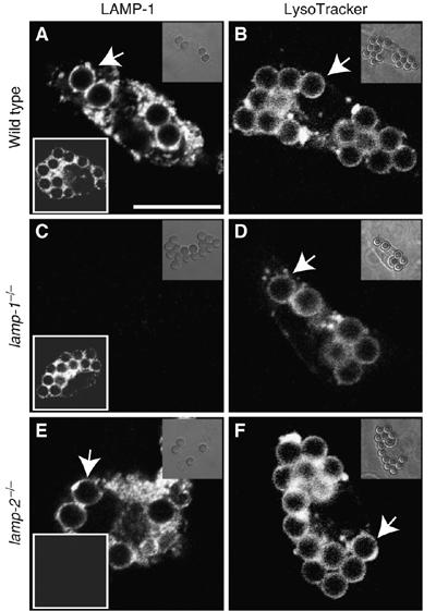
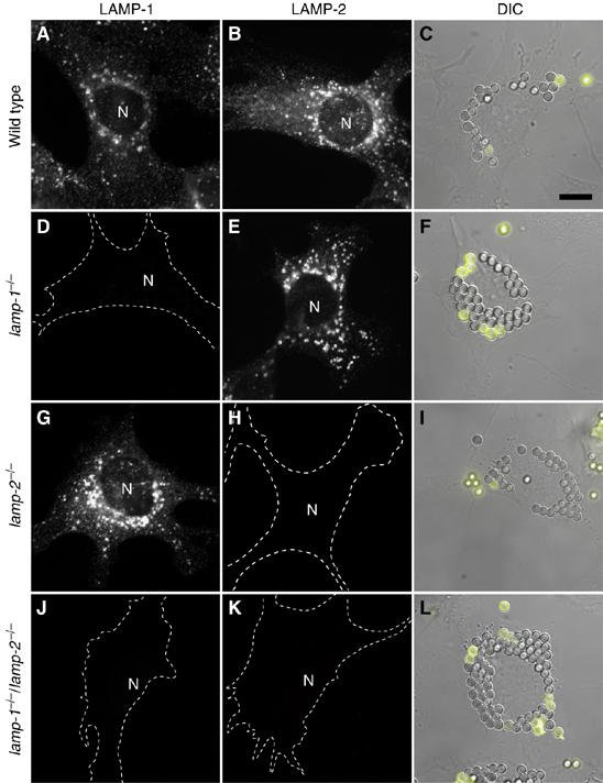
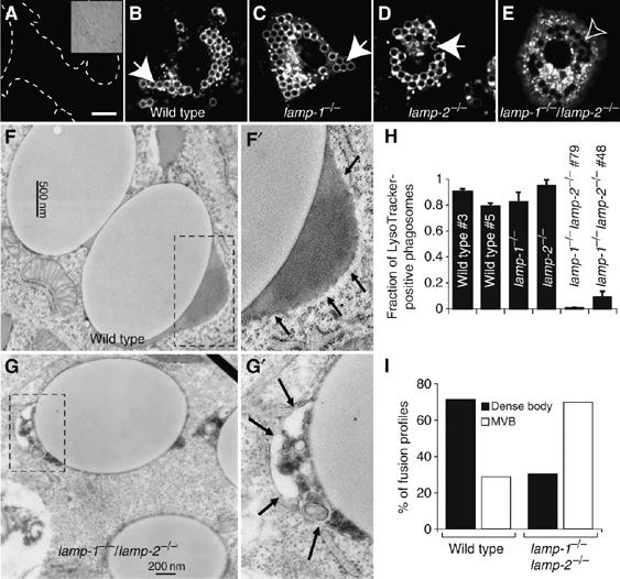
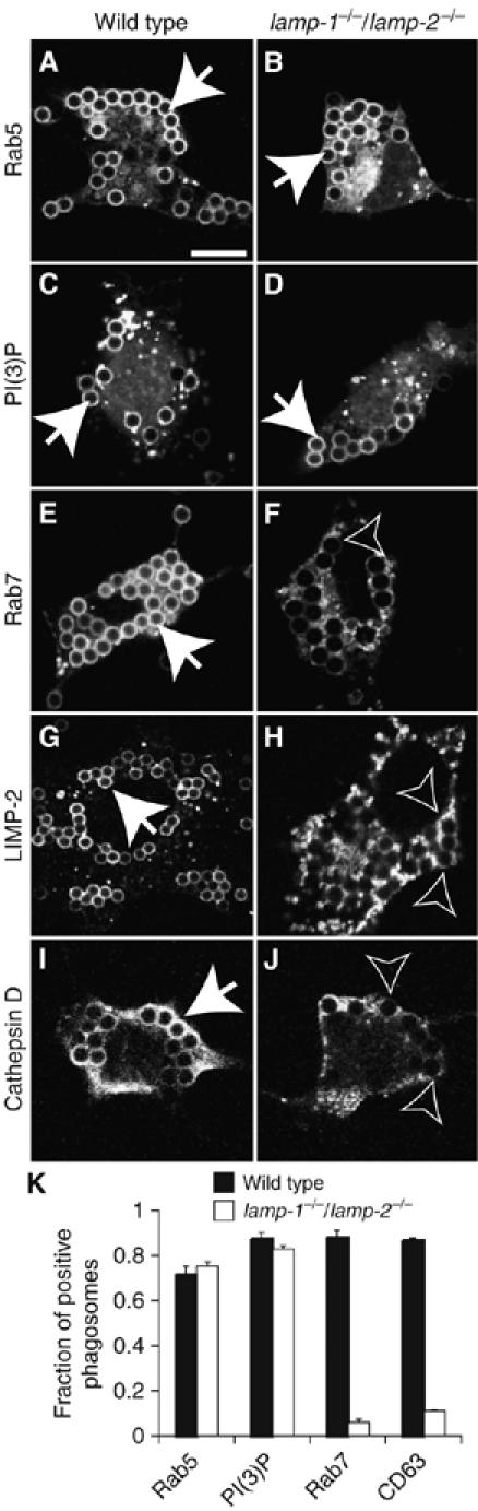
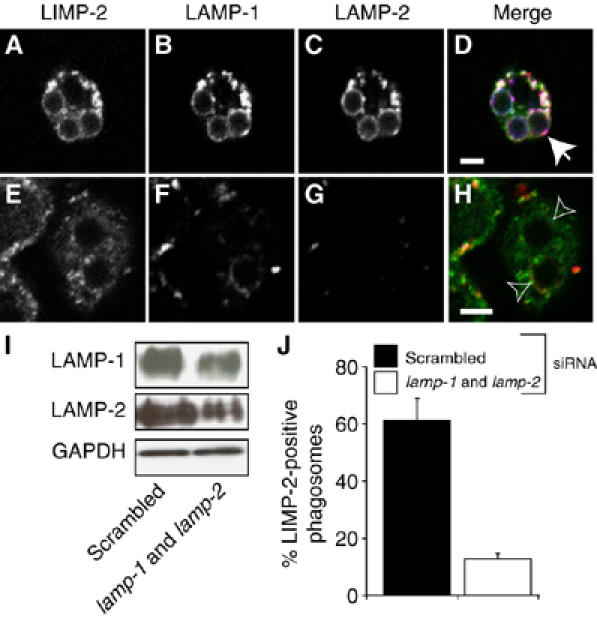
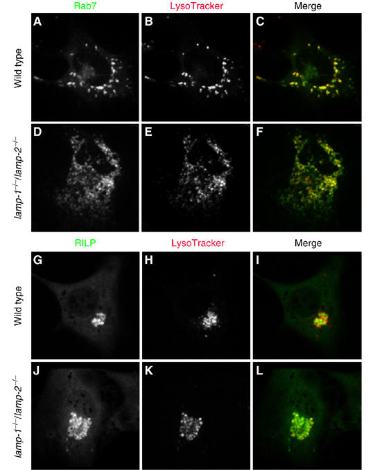
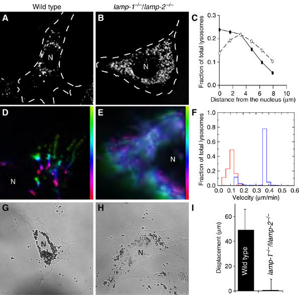
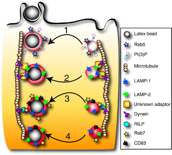
Lysosome-associated membrane proteins 1 and 2 (LAMP-1 and LAMP-2) are delivered to phagosomes during the maturation process. We used cells from LAMP-deficient mice to analyze the role of these proteins in phagosome maturation. Macrophages from LAMP-1- or LAMP-2-deficient mice displayed normal fusion of lysosomes with phagosomes. Because ablation of both the lamp-1 and lamp-2 genes yields an embryonic-lethal phenotype, we were unable to study macrophages from double knockouts. Instead, we reconstituted phagocytosis in murine embryonic fibroblasts (MEFs) by transfection of FcgammaIIA receptors. Phagosomes formed by FcgammaIIA-transfected MEFs obtained from LAMP-1- or LAMP-2- deficient mice acquired lysosomal markers. Remarkably, although FcgammaIIA-transfected MEFs from double-deficient mice ingested particles normally, phagosomal maturation was arrested. LAMP-1 and LAMP-2 double-deficient phagosomes acquired Rab5 and accumulated phosphatidylinositol 3-phosphate, but failed to recruit Rab7 and did not fuse with lysosomes. We attribute the deficiency to impaired organellar motility along microtubules. Time-lapse cinematography revealed that late endosomes/lysosomes as well as phagosomes lacking LAMP-1 and LAMP-2 had reduced ability to move toward the microtubule-organizing center, likely precluding their interaction with each other.


Figures

Figure 1
Figure 1
Phagocytosis and phagosome maturation by primary macrophages. Macrophages isolated from the peritoneum of WT (A, B), lamp-1−/− (C, D) and lamp-2−/− mice (E, F) were exposed to IgG-opsonized beads. Maturation was allowed to proceed for 60 min and macrophages were immunostained for LAMP-1 (main panels in A, C, E) and LAMP-2 (lower left insets in A, C, E) or labeled with LysoTracker (B, D, F). Arrows indicate LysoTracker-enriched phagosomes. Images are representative of at least three separate experiments of each type. Scale bar=10 μm.
Figure 2
Figure 2
LAMP immunostaining and phagocytosis by Fc receptor-transfected MEFs. MEFs from WT (AC), lamp-1−/− (DF), lamp-2−/− (GI) and lamp-1−/−/lamp-2−/− (JL) mice were stained for LAMP-1 (A, D, G, J) or LAMP-2 (B, E, H, K). Cell outlines were traced when immunostaining was negative. N=nucleus. WT (C), lamp-1−/− (F), lamp-2−/− (I) and lamp-1−/−/lamp-2−/− fibroblasts (L) were transfected with FcR. After 24 h, transfectants were exposed to beads and phagocytosis allowed to proceed for 15 min. External beads were identified by the addition of anti-IgG antibodies (yellow). Images are representative of at least 3 experiments of each type. Scale bar=10 μm.
Figure 3
Figure 3
Phagosome maturation in MEFs expressing FcγIIA receptors. (A) WT fibroblasts were treated with 10 mM NH4Cl and 250 nM concanamycin A and labeled with LysoTracker. Dashed lines show cell outline. Inset: corresponding DIC. WT (B, F), lamp-1−/− (C), lamp-2−/− (D) and lamp-1−/−/lamp-2−/− MEFs (E, G) were transfected with FcR. After 16–24 h, transfectants were exposed to beads, and after 15 min of engulfment, phagosomes were allowed to undergo maturation for 60 min. In (A–E), LysoTracker was added and cells were visualized by confocal microscopy. Closed arrows: LysoTracker-positive phagosome; open arrows: LysoTracker-negative phagosomes. Scale bar=10 μm. WT (F, F′) and lamp-1−/−/lamp-2−/− (G, G′) phagosomes were analyzed by EM. Dashed boxes were enlarged and are shown in panels F′ and G′. Arrows point to the region of interest. (H) Quantitation of the fraction of LysoTracker-positive phagosomes in WT and lamp1−/−/lamp2−/− (two clones each) and lamp1−/− and lamp2−/− MEFs. Accumulation or absence of LysoTracker around internalized latex beads was determined by confocal microscopy. Data are means±s.e. of four experiments (at least 100 phagosomes from ⩾20 cells per experiment. (I) Quantitation of phagosomes undergoing fusion with dense bodies (lysosomes, black bars) or multivesicular bodies (MVB, open bars) in WT and lamp-1−/−/lamp-2−/− MEFs. Dense body indicates fusion partners that contained homogeneous electron-dense material. MVB indicates fusion partners that contained internal membranes, mostly small vesicles. 230 and 221 phagosome fusion profiles were scored for WT and lamp-1−/−/lamp-2−/− fibroblasts, respectively.
Figure 4
Figure 4
Acquisition of endocytic markers. WT (A, C, E, G, I) and lamp-1−/−/lamp-2−/− MEFs (B, D, F, H, J) were transfected with FcR and after 24 h were exposed to IgG-opsonized beads. Phagosome maturation was allowed to proceed for 5 min in WT and lamp1−/−/lamp2−/− fibroblasts expressing GFP-Rab5 (A, B, respectively) or GFP-PX (C, D, respectively). Alternatively, maturation was allowed to proceed for 60 min in WT and lamp1−/−/lamp2−/− MEFs expressing GFP-Rab7 (E, F). WT and lamp-1−/−/lamp-2−/− MEFs were stained for LIMP-2 (G and H, respectively) and cathepsin D (I, J). (K) Quantitation of the fraction of phagosomes positive for endocytic markers in WT (black bars) and lamp1−/−/lamp2−/− fibroblasts (open bars). Data are means±s.e. of four experiments with ⩾100 phagosomes from ⩾20 different cells. Closed arrows: phagosomes positive for the indicated marker; open arrows: phagosomes negative for the markers. Scale bar=10 μm.
Figure 5
Figure 5
Phagosome maturation in macrophages treated with siRNA against lamp-1 and lamp-2. RAW 264.7 macrophages were transfected with either scrambled siRNA (A–D, left lane in I) or siRNA against lamp-1 and lamp-2 (E–H, right lane in I). (A–H) Macrophages treated with scrambled (A–D) or lamp-1 and lamp-2 siRNA (E, F) were exposed to beads and maturation allowed to proceed for 60 min. Cells were stained for LIMP-2 (A, E; green in D) to assess phagolysosome fusion and for LAMP-1 (B, F; red in D) and LAMP-2 (C, G; blue in D) to determine knockdown effectiveness. Merged images are shown in (D) and (H). Closed arrows: phagosomes enriched with LIMP-2; open arrows: phagosomes negative for LIMP-2. (I) Whole-cell lysates were probed by immunoblotting for LAMP-1 and LAMP-2 72 h later. Blots were stripped and reprobed for GAPDH as loading control. (J) Quantitation of the effect of LAMP-1 and LAMP-2 knockdown on phagolysosome biogenesis. Phagolysosomes were identified by LIMP-2 staining. Data represent means±s.e. from three experiments with ⩾30 phagosomes from ⩾10 cells. Scale bar=3 μm.
Figure 6
Figure 6
Colocalization of Rab7 and RILP with LysoTracker-labeled compartments. WT (A–C, G–I) and lamp-1−/−/lamp-2−/− fibroblasts (D–F, J–L) were transfected with cDNA encoding GFP-Rab7 (A–F) or GFP-RILP (G–L). After 24 h, acidic compartments were labeled with LysoTracker and the cells visualized by confocal microscopy. Rab7 distribution is shown in (A) and (D); RILP distribution is shown in (G) and (J); LysoTracker is shown in (B), (E), (H) and (K); merged images are shown in (C), (F), (I) and (L). Images are representative of ⩾3 experiments.
Figure 7
Figure 7
Determination of lysosome and phagosome motility. Acidic compartments of WT (A, D) and lamp-1−/−/lamp-2−/− MEFs (B, E) were loaded with LysoTracker. Their distribution at steady state was recorded by epifluorescence microscopy (A, B) and the cell outline, determined by DIC, is shown by dotted line. In (C), the distribution of individual lysosomes at rest was determined relative to the nucleus using the Cellomics system. The distribution of lysosomes, expressed as a fraction of the total, is plotted as a function of distance (μm) from the nucleus in WT (solid symbols) and lamp-1−/−/lamp-2−/− cells (open symbols). Data are means of three experiments, scoring ⩾500 cells per experiment. The error bars (±s.e.) were smaller than the symbol and are not visible. In (D) and (E), the response of lysosomes to cytosolic alkalinization was documented using time-lapse microscopy. The path of a given lysosome is shown by a pseudocolor scale (vertical color bar) where yellow-green indicates the starting position and red the final position attained 10 min after alkalinization. White indicates lysosomes that did not move. (F) Summary of automated determinations of lysosomal velocities. Late endosome/lysosomes of WT (blue) and lamp-1−/−/lamp-2−/− (red) were labeled with LysoTracker. Retrograde transport upon alkalinization of the cytosol was determined for a period of 10 min as described in Materials and Methods. WT (G) and lamp-1−/−/lamp-2−/− cells (H) were exposed to 0.8 μm beads and after 3 h, their distribution was imaged by DIC. (I) Quantitation of displacement of internalized beads from the plasma membrane over 30 min. Data are means+s.e. of three experiments, scoring ⩾18 phagosomes per cell. N=nucleus.
Figure 8
Figure 8
Schematic representation of the proposed role of LAMPs in maturation. Following sealing, the phagosome (left vertical path) is propelled into the cell where a series of successive fusion events with endocytic vesicles (right vertical sequence) occurs. Phagosome maturation begins with fusion to early endosomes (1) that coincides with acquisition of PI(3)P and Rab5. These are needed for subsequent interactions with late endosomes. We propose that LAMP-1 and LAMP-2 can be acquired independently and before Rab7 (2). LAMP isoforms promote docking of phagosomes onto microtubules and centripetal movement (downward) by association with dynein/dynactin directly or via an unknown adaptor. LAMPs then mediate fusion with traditional late endosomes carrying Rab7 (3). Maturation culminates with phagosome-lysosome fusion (4), forming a microbicidal organelle.

References

    1. Andrejewski N, Punnonen EL, Guhde G, Tanaka Y, Lullmann-Rauch R, Hartmann D, von Figura K, Saftig P (1999) Normal lysosomal morphology and function in LAMP-1-deficient mice. J Biol Chem 274: 12692–12701 - PubMed
    1. Blocker A, Griffiths G, Olivo JC, Hyman AA, Severin FF (1998) A role for microtubule dynamics in phagosome movement. J Cell Sci 111 (Part 3): 303–312 - PubMed
    1. Bucci C, Thomsen P, Nicoziani P, McCarthy J, van Deurs B (2000) Rab7: a key to lysosome biogenesis. Mol Biol Cell 11: 467–480 - PMC - PubMed
    1. Cantalupo G, Alifano P, Roberti V, Bruni CB, Bucci C (2001) Rab-interacting lysosomal protein (RILP): the Rab7 effector required for transport to lysosomes. EMBO J 20: 683–693 - PMC - PubMed
    1. Chavrier P, Parton RG, Hauri HP, Simons K, Zerial M (1990) Localization of low molecular weight GTP binding proteins to exocytic and endocytic compartments. Cell 62: 317–329 - PubMed

Publication types

MeSH terms

Substances