

Highly potent triazole-based tubulin polymerization inhibitors
- PMID: 17249649
- PMCID: PMC2694353
- DOI: 10.1021/jm061142s
Highly potent triazole-based tubulin polymerization inhibitors
Abstract
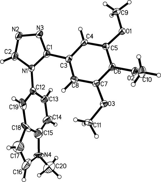
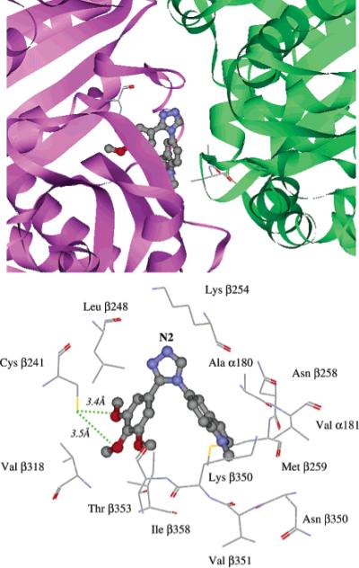
We describe the synthesis and biological evaluation of a series of tubulin polymerization inhibitors that contain the 1,2,4-triazole ring to retain the bioactive configuration afforded by the cis double bond in combretastatin A-4 (CA-4). Several of the subject compounds exhibited potent tubulin polymerization inhibitory activity as well as cytotoxicity against a variety of cancer cells including multi-drug-resistant (MDR) cancer cell lines. Attachment of the N-methyl-5-indolyl moiety to the 1,2,4-triazole core, as exemplified by compound 7, conferred optimal properties among this series. Computer docking and molecular simulations of 7 inside the colchicine binding site of tubulin enabled identification of residues most likely to interact strongly with these inhibitors and explain their potent anti-tubulin activity and cytotoxicity. It is hoped that results presented here will stimulate further examination of these substituted 1,2,4-triazoles as potential anti-cancer therapeutic agents.
Figures


References
-
- Ducki S, Mackenzie G, Lawrence NJ, Snyder JP. Quantitative structure–activity relationship (5D-QSAR) study of combretastatin-like analogues as inhibitors of tubulin assembly. J. Med. Chem. 2005;48:457–465. - PubMed
-
- Jordan A, Hadfield JA, Lawrence NJ, McGown AT. Tubulin as a target for anticancer drugs: agents that interact with the mitotic spindle. Med. Res. Rev. 1998;18:259–296. - PubMed
-
- Andreu JM, Barasoain I. The interaction of baccatin III with the taxol binding site of microtubules determined by a homogeneous assay with fluorescent taxoid. Biochemistry. 2001;40:11975–11984. - PubMed
-
- Rai SS, Wolff J. Localization of the vinblastine-binding site on beta-tubulin. J. Biol. Chem. 1996;271:14707–14711. - PubMed
-
- ter Haar E, Rosenkranz HS, Hamel E, Day BW. Computational and molecular modeling evaluation of the structural basis for tubulin polymerization inhibition by colchicine site agents. Bioorg. Med. Chem. 1996;4:1659–1671. - PubMed
Publication types
MeSH terms
Substances
Grants and funding
LinkOut - more resources
Full Text Sources
Other Literature Sources
