

A novel superfamily containing the beta-grasp fold involved in binding diverse soluble ligands
- PMID: 17250770
- PMCID: PMC1796856
- DOI: 10.1186/1745-6150-2-4
A novel superfamily containing the beta-grasp fold involved in binding diverse soluble ligands
Abstract
Background: Domains containing the beta-grasp fold are utilized in a great diversity of physiological functions but their role, if any, in soluble or small molecule ligand recognition is poorly studied.
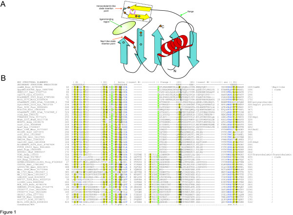
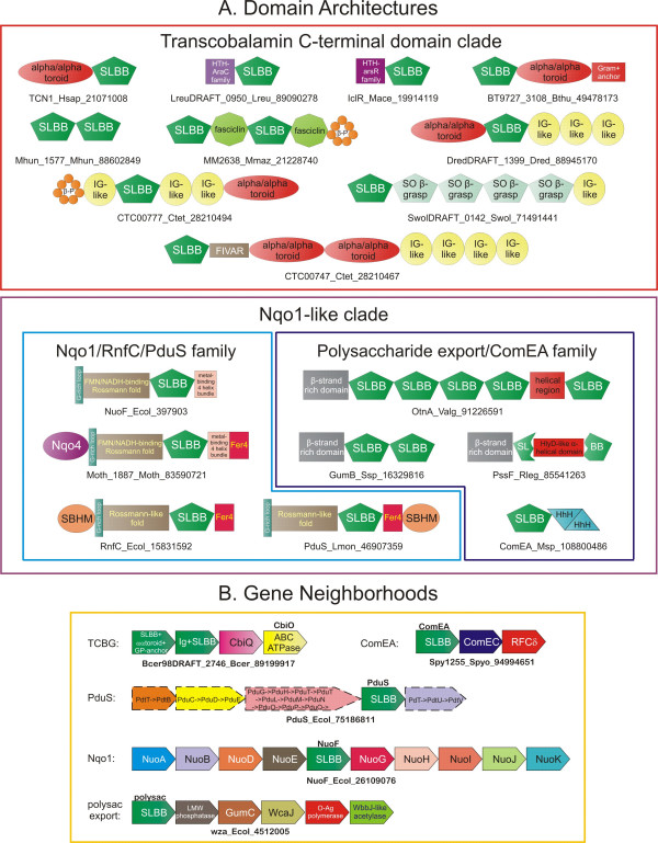
Results: Using sensitive sequence and structure similarity searches we identify a novel superfamily containing the beta-grasp fold. They are found in a diverse set of proteins that include the animal vitamin B12 uptake proteins transcobalamin and intrinsic factor, the bacterial polysaccharide export proteins, the competence DNA receptor ComEA, the cob(I)alamin generating enzyme PduS and the Nqo1 subunit of the respiratory electron transport chain. We present evidence that members of this superfamily are likely to bind a range of soluble ligands, including B12. There are two major clades within this superfamily, namely the transcobalamin-like clade and the Nqo1-like clade. The former clade is typified by an insert of a beta-hairpin after the helix of the beta-grasp fold, whereas the latter clade is characterized by an insert between strands 4 and 5 of the core fold.
Conclusion: Members of both clades within this superfamily are predicted to interact with ligands in a similar spatial location, with their specific inserts playing a role in the process. Both clades are widely represented in bacteria suggesting that this superfamily was derived early in bacterial evolution. The animal lineage appears to have acquired the transcobalamin-like proteins from low GC Gram-positive bacteria, and this might be correlated with the emergence of the ability to utilize B12 produced by gut bacteria.
Reviewers: This article was reviewed by Andrei Osterman, Igor Zhulin, and Arcady Mushegian.
Figures
Similar articles
-
Vitamin B12 transport proteins: crystallographic analysis of beta-axial ligand substitutions in cobalamin bound to transcobalamin.IUBMB Life. 2007 Nov;59(11):722-9. doi: 10.1080/15216540701673413. IUBMB Life. 2007. PMID: 17943552
-
Protein signatures (molecular synapomorphies) that are distinctive characteristics of the major cyanobacterial clades.Int J Syst Evol Microbiol. 2009 Oct;59(Pt 10):2510-26. doi: 10.1099/ijs.0.005678-0. Epub 2009 Jul 21. Int J Syst Evol Microbiol. 2009. PMID: 19622649
-
RCC1-like repeat proteins: a pangenomic, structurally diverse new superfamily of beta-propeller domains.Proteins. 2008 Feb 1;70(2):378-87. doi: 10.1002/prot.21521. Proteins. 2008. PMID: 17680689
-
Safety and nutritional assessment of GM plants and derived food and feed: the role of animal feeding trials.Food Chem Toxicol. 2008 Mar;46 Suppl 1:S2-70. doi: 10.1016/j.fct.2008.02.008. Epub 2008 Feb 13. Food Chem Toxicol. 2008. PMID: 18328408 Review.
-
Relaxin-3, INSL5, and their receptors.Results Probl Cell Differ. 2008;46:213-37. doi: 10.1007/400_2007_055. Results Probl Cell Differ. 2008. PMID: 18236022 Review.
Cited by
-
Ionic strength-dependent conformations of a ubiquitin-like small archaeal modifier protein (SAMP2) from Haloferax volcanii.Sci Rep. 2013;3:2136. doi: 10.1038/srep02136. Sci Rep. 2013. PMID: 23823798 Free PMC article.
-
Small but versatile: the extraordinary functional and structural diversity of the beta-grasp fold.Biol Direct. 2007 Jul 2;2:18. doi: 10.1186/1745-6150-2-18. Biol Direct. 2007. PMID: 17605815 Free PMC article.
-
Extending the aerolysin family: from bacteria to vertebrates.PLoS One. 2011;6(6):e20349. doi: 10.1371/journal.pone.0020349. Epub 2011 Jun 8. PLoS One. 2011. PMID: 21687664 Free PMC article.
-
The natural history of ubiquitin and ubiquitin-related domains.Front Biosci (Landmark Ed). 2012 Jan 1;17(4):1433-60. doi: 10.2741/3996. Front Biosci (Landmark Ed). 2012. PMID: 22201813 Free PMC article. Review.
-
Regulation of BRCA1 stability through the tandem UBX domains of isoleucyl-tRNA synthetase 1.Nat Commun. 2022 Nov 8;13(1):6732. doi: 10.1038/s41467-022-34612-y. Nat Commun. 2022. PMID: 36347866 Free PMC article.
References
-
- Wolf YI, Aravind L, Grishin NV, Koonin EV. Evolution of aminoacyl-tRNA synthetases--analysis of unique domain architectures and phylogenetic trees reveals a complex history of horizontal gene transfer events. Genome Res. 1999;9:689–710. - PubMed
-
- Chishti AH, Kim AC, Marfatia SM, Lutchman M, Hanspal M, Jindal H, Liu SC, Low PS, Rouleau GA, Mohandas N, Chasis JA, Conboy JG, Gascard P, Takakuwa Y, Huang SC, Benz EJ, Jr., Bretscher A, Fehon RG, Gusella JF, Ramesh V, Solomon F, Marchesi VT, Tsukita S, Tsukita S, Hoover KB, et al. The FERM domain: a unique module involved in the linkage of cytoplasmic proteins to the membrane. Trends Biochem Sci. 1998;23:281–282. doi: 10.1016/S0968-0004(98)01237-7. - DOI - PubMed
LinkOut - more resources
Full Text Sources
Miscellaneous
