

Spironolactone improves structure and increases tone in the cerebral vasculature of male spontaneously hypertensive stroke-prone rats
- PMID: 17250855
- PMCID: PMC1913209
- DOI: 10.1016/j.mvr.2006.12.001
Spironolactone improves structure and increases tone in the cerebral vasculature of male spontaneously hypertensive stroke-prone rats
Abstract
Background: Previous studies show that ischemic cerebral infarct size is related to cerebral vessel structure. Spironolactone, a mineralocorticoid receptor antagonist, decreases ischemic cerebral infarct size in male spontaneously hypertensive stroke-prone rats (SHRSP). Therefore, we hypothesized that chronic spironolactone treatment would improve cerebral artery structure in the SHRSP.
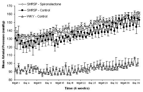
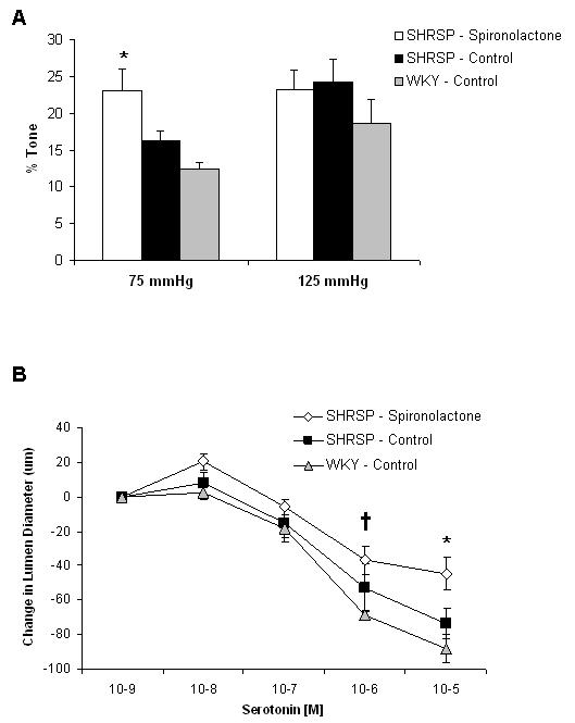
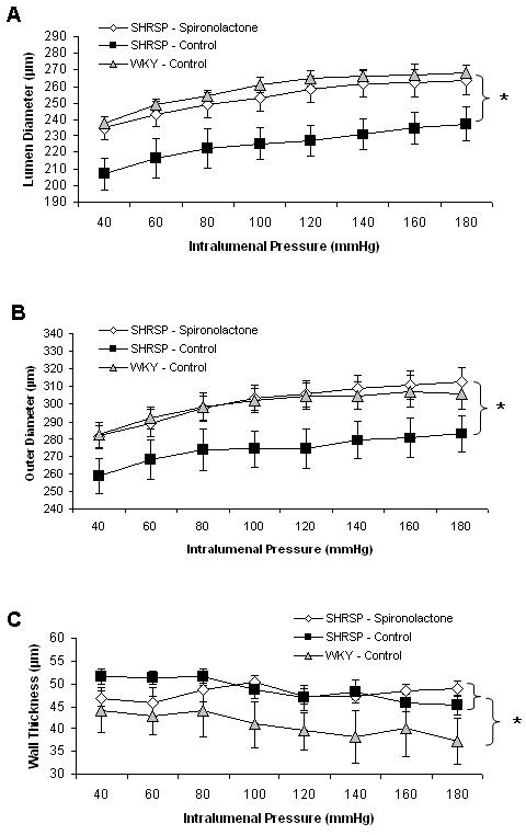
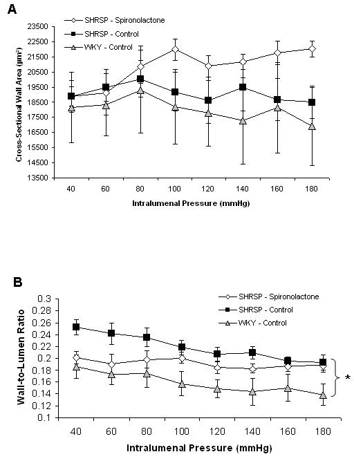
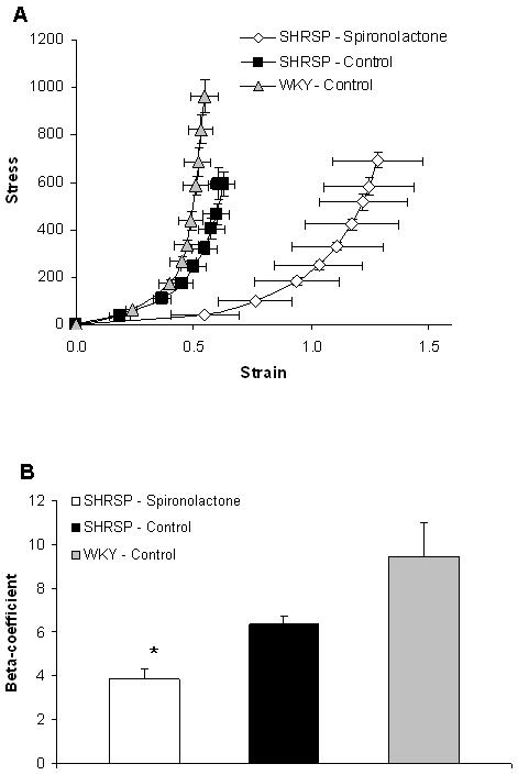
Methods: Six-week-old male SHRSP were treated with spironolactone (2.5 mg/day) for 6 weeks and were compared to untreated control SHRSP and normotensive Wistar Kyoto (WKY) rats. Using a pressurized arteriograph, structural measurements of the middle cerebral artery (MCA) were taken under passive (calcium-free), zero-flow conditions. Myogenic tone was calculated from active and passive measurements taken at 75 and 125 mmHg. Mean arterial pressure was measured using radiotelemetry.
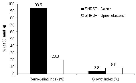
Results: Myogenic tone was increased only at 75 mmHg in the spironolactone-treated SHRSP compared to control rats. The MCA lumen and outer diameters were increased in the spironolactone-treated SHRSP compared to control SHRSP, but were not different from WKY rats, indicating a decrease in vascular remodeling. There was no effect of spironolactone on blood pressure, suggesting that this is a blood pressure-independent effect.
Conclusion: Increased myogenic tone and lumen diameter in the spironolactone-treated SHRSP may be responsible for the protective role of spironolactone in ischemic stroke.
Figures
Similar articles
-
Intact female stroke-prone hypertensive rats lack responsiveness to mineralocorticoid receptor antagonists.Am J Physiol Regul Integr Comp Physiol. 2007 Oct;293(4):R1754-63. doi: 10.1152/ajpregu.00145.2007. Epub 2007 Aug 1. Am J Physiol Regul Integr Comp Physiol. 2007. PMID: 17670862 Free PMC article.
-
Regulation of myogenic tone and structure of parenchymal arterioles by hypertension and the mineralocorticoid receptor.Am J Physiol Heart Circ Physiol. 2015 Jul 1;309(1):H127-36. doi: 10.1152/ajpheart.00168.2015. Epub 2015 Apr 24. Am J Physiol Heart Circ Physiol. 2015. PMID: 25910805 Free PMC article.
-
Effects of spironolactone on cerebral vessel structure in rats with sustained hypertension.Am J Hypertens. 2011 Jun;24(6):708-15. doi: 10.1038/ajh.2011.20. Epub 2011 Feb 24. Am J Hypertens. 2011. PMID: 21350432 Free PMC article.
-
Pathological alterations of astrocytes in stroke-prone spontaneously hypertensive rats under ischemic conditions.Neurochem Int. 2012 Jan;60(1):91-8. doi: 10.1016/j.neuint.2011.11.002. Epub 2011 Nov 15. Neurochem Int. 2012. PMID: 22100568 Review.
-
[Comparison of biochemical properties and protein level of mevalonate pyrophosphate decarboxylase between stroke-prone spontaneously hypertensive rats and Wistar-Kyoto rats].Yakugaku Zasshi. 2004 Oct;124(10):683-92. doi: 10.1248/yakushi.124.683. Yakugaku Zasshi. 2004. PMID: 15467276 Review. Japanese.
Cited by
-
Improvement in middle cerebral artery structure and endothelial function in stroke-prone spontaneously hypertensive rats after macrophage depletion.Microcirculation. 2013 Oct;20(7):650-61. doi: 10.1111/micc.12064. Microcirculation. 2013. PMID: 23647512 Free PMC article.
-
Multimodal markers of inflammation in the subcortical ischemic vascular disease type of vascular cognitive impairment.Stroke. 2014 May;45(5):1531-8. doi: 10.1161/STROKEAHA.113.004534. Epub 2014 Apr 1. Stroke. 2014. PMID: 24692476 Free PMC article. Review. No abstract available.
-
Intact female stroke-prone hypertensive rats lack responsiveness to mineralocorticoid receptor antagonists.Am J Physiol Regul Integr Comp Physiol. 2007 Oct;293(4):R1754-63. doi: 10.1152/ajpregu.00145.2007. Epub 2007 Aug 1. Am J Physiol Regul Integr Comp Physiol. 2007. PMID: 17670862 Free PMC article.
-
Impaired myogenic constriction of the renal afferent arteriole in a mouse model of reduced βENaC expression.Am J Physiol Renal Physiol. 2012 Jun 1;302(11):F1486-93. doi: 10.1152/ajprenal.00638.2011. Epub 2012 Mar 14. Am J Physiol Renal Physiol. 2012. PMID: 22419697 Free PMC article.
-
The development of hypertension and hyperaldosteronism in a rodent model of life-long obesity.Endocrinology. 2012 Apr;153(4):1764-73. doi: 10.1210/en.2011-1176. Epub 2012 Feb 21. Endocrinology. 2012. PMID: 22355066 Free PMC article.
References
-
- Baumbach GL, Heistad DD. Remodeling of cerebral arterioles in chronic hypertension. Hypertension. 1989;13:968–972. - PubMed
-
- Benetos A, Lacolley P, Safar ME. Prevention of aortic fibrosis by spironolactone in spontaneously hypertensive rats. Arterioscler Thromb Vasc Biol. 1997;17:1152–1156. - PubMed
-
- Brunner HR, Laragh JH, Baer L, Newton MA, Goodwin FT, Krakoff LR, Bard RH, Buhler FR. Essential hypertension: renin and aldosterone, heart attack and stroke. N Engl J Med. 1972;286:441–449. - PubMed
-
- Cipolla MJ, Lessov N, Clark WM, Haley EC., Jr. Postischemic attenuation of cerebral artery reactivity is increased in the presence of tissue plasminogen activator. Stroke. 2000;31:940–945. - PubMed
-
- Cipolla MJ, Lessov N, Hammer ES, Curry AB. Threshold Duration of Ischemia for Myogenic Tone in Middle Cerebral Arteries : Effect on Vascular Smooth Muscle Actin. Stroke. 2001;32:1658–1664. - PubMed
Publication types
MeSH terms
Substances
Grants and funding
LinkOut - more resources
Full Text Sources
Medical
