. 2007 Apr;81(8):4145-57.
doi: 10.1128/JVI.02685-06. Epub 2007 Jan 24.

Sensing infection by adenovirus: Toll-like receptor-independent viral DNA recognition signals activation of the interferon regulatory factor 3 master regulator

Affiliations

Sensing infection by adenovirus: Toll-like receptor-independent viral DNA recognition signals activation of the interferon regulatory factor 3 master regulator

Marcelo Nociari et al. J Virol. 2007 Apr.

Abstract

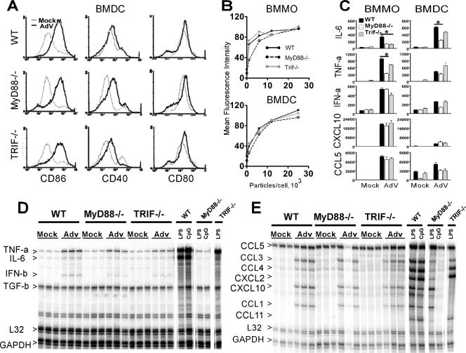
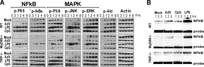
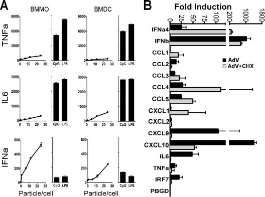
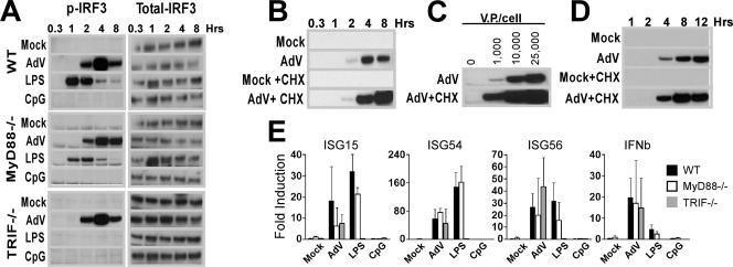
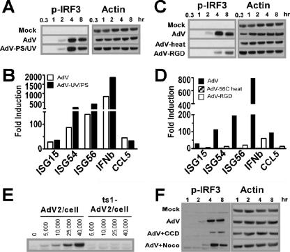
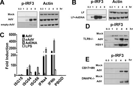
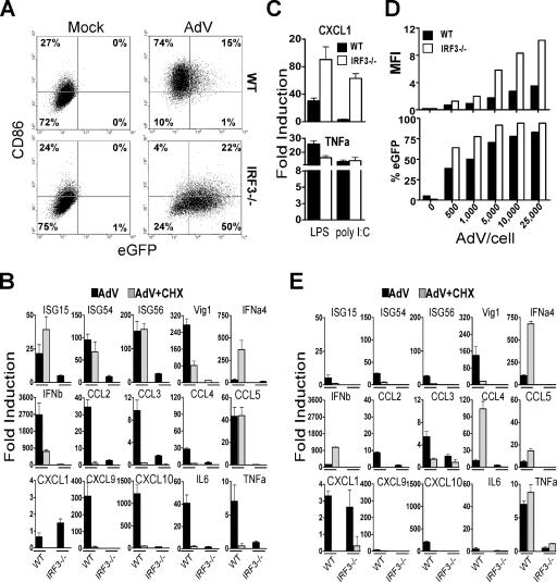
Infection with adenovirus vectors (AdV) results in rapid activation of innate immunity, which serves the dual purpose of stimulating inflammatory antiviral host defenses and the adaptive immune system. Viral recognition by macrophages, dendritic cells, and other cell types requires an ability to sense the presence of a foreign molecular pattern by "pattern recognition receptors." The nature of the adenoviral sensor, the target ligand of the sensor, and the downstream antiviral signaling response triggered by virus infection have not been defined for this nonenveloped double-stranded DNA (dsDNA) virus. We have identified four critical links involved in AdV recognition by murine antigen-presenting cells (APC) and primary lung fibroblasts: (i) viral recognition occurs chiefly via a Toll-like receptor (TLR)-independent nucleic acid-sensing mechanism recognizing the viral dsDNA genome, (ii) the intact viral particle and capsid proteins are required for efficient intracellular delivery of the viral genome, (iii) delivery of the viral genome triggers interferon regulatory factor 3 (IRF3) phosphorylation, and (iv) IRF3 activation is the required dominant antiviral signaling pathway used by APC, whereas the "primary" involvement of NF-kappaB, mitogen-activated protein kinase, or Akt pathways is less prominent. In this study we provide the first direct evidence that infection by a dsDNA virus stimulates an IRF3-mediated interferon and proinflammatory response through a TLR-independent DNA-sensing mechanism.


Figures

FIG. 1.
FIG. 1.
Role of MyD88 and TRIF in the innate immune response to AdV. (A) Expression of CD86, CD40, and CD80 on gated CD11c+ BMDC after 36 h of treatment with 5,000 AdV particles/cell (black) or mock treatment (gray). (B) CD86 expression in BMMO and BMDC from WT, MyD88−/−, and TRIF−/− mice incubated with various doses of AdV at 36 h postinfection. (C) Induction of cytokines in macrophages and DC derived from WT, MyD88−/−, and TRIF−/− mice. Cells were incubated with 25,000 AdV particles/cell for 36 h, and supernatant cytokine levels (pg/ml) were determined by ELISA. Error bars represent standard deviations for triplicate wells in a single experiment; data are representative of four independent experiments. Student's t tests were completed, and P values of <0.05 are indicated (*). (D and E) RPA analysis for cytokines (D) and chemokines (E) of total RNA isolated from BMDC of the indicated genotypes after 5 h of treatment with 25,000 AdV particles/cell, 10 ng/ml LPS, or 2 μM CpG DNA. The triplicate results shown for AdV are from independent infections and are representative of two separate experiments. GAPDH, glyceraldehyde-3-phosphate dehydrogenase.
FIG. 2.
FIG. 2.
Biochemical analysis of signaling responses to AdV, MyD88, and MyD88/TRIF agonists. (A) Western blot analysis of signal transduction using the indicated antibodies. At each indicated time point, whole-cell lysates from mock-treated macrophages were compared to lysates from macrophages stimulated with either 25,000 AdV particles/cell, 10 ng/ml LPS, or 2 μM CpG DNA. Data are representative of three independent experiments. (B) NF-κB gel shift assay. Nuclear extracts from macrophages stimulated for the indicated times were used in gel shift assay with an [α-32P]ATP-labeled probe containing a consensus NF-κB-binding sequence.
FIG. 3.
FIG. 3.
AdV induce a primary type I IFN and chemokine response in APC. (A) Cytokines produced by BMMO and BMDC after stimulation for 36 h with various doses of AdV, 10 ng/ml LPS, or 2 μM CpG DNA. (B) QRT-PCR analysis of type I IFNs and proinflammatory cytokines induced in BMMO after 5 h of treatment with buffer or 25,000 AdV particles/cell. White and black bars represent the fold induction in the absence and in the presence of 50 μg/ml CHX, respectively. Cell treatments were performed in triplicate. β-Actin levels were quantified in all total RNA samples for normalization of the data. The fold change in gene expression was determined by comparison to the mock-treated samples. IRF7 was monitored to assess CHX effectiveness, and the porphobilinogen deaminase (PBGD) housekeeping gene was used as negative control. Two independent real-time PCR experiments gave comparable results.
FIG. 4.
FIG. 4.
AdV activate an IRF3-mediated innate immune response. (A to D) Ser396P-IRF3 levels in whole-cell lysates from BMMO. AdV stimulations were performed at 25,000 viral particles per cell unless otherwise indicated. (A) WT, MyD88-KO, and TRIF-KO BMMO were stimulated with buffer, AdV, 10 ng/ml LPS, or 2 μM CpG DNA for the indicated times. After Ser396P-IRF3 determination, membranes were stripped and reprobed using a pan-IRF3 specific antibody. (B) WT macrophages were stimulated with buffer or AdV with and without CHX; whole-cell lysates were prepared at the indicated times. (C) Macrophages were stimulated with various doses of AdV in the presence or absence of CHX for 4 h. (D) IRF3 phosphorylation from primary lung fibroblasts treated with or without 25,000 particles AdV/cell with or without CHX over a 12-h time course. (E) Induction of IRF3-dependent gene expression in BMMO. Total RNA from BMMO infected at 25,000 AdV particles/cell for 5 h in the presence of CHX (to avoid ISGF3 gene induction) was analyzed by QRT-PCR. Cell treatments were performed in triplicate. β-Actin levels were quantified for normalization of the data. The fold change in gene expression was determined by comparison to the mock-treated samples. Data are representative of at least three independent experiments.
FIG. 5.
FIG. 5.
Viral entrance but not viral transcription or nuclear localization is required for AdV activation of IRF3. (A) Ser396P-IRF3 and actin Western blots of lysates from macrophages stimulated with buffer, AdV, or UV/psoralen-inactivated AdV (multiplicity of infection, 25,000 particles/cell) (B) Real-time PCR analysis comparing the induction of IRF3-dependent genes by AdV (white bars) and by UV/psoralen-inactivated AdV (black bars) at a multiplicity of infection of 25,000 particles/cell. Total RNA from BMMO was harvested after 5 h. β-Actin levels were quantified for normalization of the data. The fold change in gene expression was determined by comparison to the mock-treated samples. Data are representative of at least three independent experiments. (C) Ser396P-IRF3 and actin Western blots of lysates from macrophages stimulated with buffer, AdV, heat-inactivated (1 h at 56°C) AdV, or penton base-RGD mutant AdV (AdV-RGD) at a multiplicity of infection of 25,000 particles/cell. (D) Real-time PCR analysis comparing the induction of IRF3-dependent genes by AdV (black bars), 56°C AdV (gray bars), and AdV-RGD (white bars). (E) Ser396P-IRF3 and actin Western blots of lysates from macrophages infected with 25,000 particles/cell UV/psoralen-inactivated WT Ad2 or ts1 mt Ad2 and harvested at the indicated times. (F) Ser396P-IRF3 and actin Western blots of lysates from macrophages stimulated with buffer or AdV in the presence of cytochalasin D (CCD) or nocodazole (Noco) for the indicated times. Western blot data are representative of three independent experiments.
FIG. 6.
FIG. 6.
Viral DNA and not capsid protein is the IRF3-activating ligand of Ad. (A, B, D, and E) Western blots showing levels of Ser396P-IRF3 and corresponding β-actin in lysates from BMMO that were stimulated for the times indicated. (A) Macrophages were infected with buffer, AdV, or eAdV. (B) BMMO were transfected with buffer or AdV genomic DNA. (C) Total RNA was isolated from BMMO 5 h after stimulation with AdV or eAdV or transfection with AdV DNA. β-Actin levels were quantified for normalization of the data, and the fold change in gene expression (compared to a mock-treated sample) was quantified by QRT-PCR. (D) BMMO from TLR9-defective mice were infected with buffer, AdV, or HSV-1 (multiplicity of infection, 5). (E) BMMO from SCID mice (DNA-PKcs defective) and from CB-17 controls were treated with buffer or AdV.
FIG. 7.
FIG. 7.
IRF3 Is essential for innate immune responses against AdV. (A) BMMO from WT and IRF3−/− mice were stimulated for 36 h with buffer or AdV at 25,000 particles/cell. CD86 upregulation and eGFP expression were analyzed by flow cytometry. The number in each quadrant represents the percentage of positive cells. (B) QRT-PCR analysis of total RNA isolated from macrophages derived from WT and IRF3−/− mice 5 h after stimulation with buffer or AdV at 25,000 particles/cell in the presence or absence of CHX. (C) mRNA levels of CXCL1 and TNF-α in macrophages stimulated with 10 ng/ml LPS or 10 μg poly (I·C) with Lipofectamine 2000. (D) Mean fluorescence intensity (MFI) and percentage of eGFP-positive lung fibroblasts infected for 36 h with the indicated doses of AdV. (E) QRT-PCR analysis of mRNA levels induced in lung fibroblasts derived from WT and IRF3-KO mice. The experiments were performed similarly to those for panel B except that the total RNA was harvested at 8 h after stimulation. β-Actin levels were quantified for normalization of the data. The fold change in gene expression was determined by comparison to the mock-treated samples. Data are representative of at least three independent experiments.

Similar articles

Cited by

References

    1. Bergelson, J. M., J. A. Cunningham, G. Droguett, E. A. Kurt-Jones, A. Krithivas, J. S. Hong, M. S. Horwitz, R. L. Crowell, and R. W. Finberg. 1997. Isolation of a common receptor for Coxsackie B viruses and adenoviruses 2 and 5. Science 275:1320-1323. - PubMed
    1. Bieback, K., E. Lien, I. M. Klagge, E. Avota, J. Schneider-Schaulies, W. P. Duprex, H. Wagner, C. J. Kirschning, V. Ter Meulen, and S. Schneider-Schaulies. 2002. Hemagglutinin protein of wild-type measles virus activates toll-like receptor 2 signaling. J. Virol. 76:8729-8736. - PMC - PubMed
    1. Compton, T., E. A. Kurt-Jones, K. W. Boehme, J. Belko, E. Latz, D. T. Golenbock, and R. W. Finberg. 2003. Human cytomegalovirus activates inflammatory cytokine responses via CD14 and Toll-like receptor 2. J. Virol. 77:4588-4596. - PMC - PubMed
    1. Covert, M. W., T. H. Leung, J. E. Gaston, and D. Baltimore. 2005. Achieving stability of lipopolysaccharide-induced NF-kappaB activation. Science 309:1854-1857. - PubMed
    1. Genin, P., M. Algarte, P. Roof, R. Lin, and J. Hiscott. 2000. Regulation of RANTES chemokine gene expression requires cooperativity between NF-kappa B and IFN-regulatory factor transcription factors. J. Immunol. 164:5352-5361. - PubMed

Publication types

MeSH terms

LinkOut - more resources