Review
. 2007 Feb 1;120(Pt 3):379-83.
doi: 10.1242/jcs.03349.

Metabolic catastrophe as a means to cancer cell death

Affiliations
Review

Metabolic catastrophe as a means to cancer cell death

Shengkan Jin et al. J Cell Sci. .

Abstract

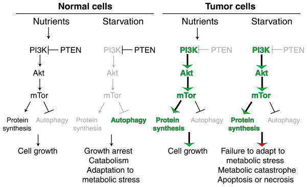
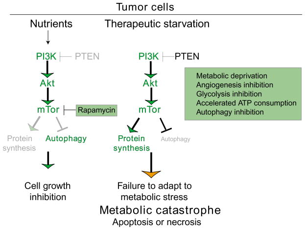
During tumorigenesis, normal growth mechanisms are deregulated and safeguards that eliminate abnormal cells by apoptosis are disabled. Tumor cells must also increase nutrient uptake and angiogenesis to support the upregulation of metabolism necessary for unrestricted growth. In addition, they have to rely on inefficient energy production by glycolysis. This glycolytic state can result from mutations that promote cell proliferation, the hypoxic tumor microenvironment and perhaps mitochondrial malfunction. Moreover, the very signals that enable unrestricted cell proliferation inhibit autophagy, which normally sustains cells during nutrient limitation. In tumors, inactivation of the autophagy pathway may enhance necrosis and inflammation and promote genomic instability, which can further enhance tumor growth. Thus, tumor cells cannot adapt efficiently to metabolic stress and could be induced to die by metabolic catastrophe, in which high energy demand is contrasted by insufficient energy production. Efforts to exploit this unique metabolic state clinically previously focused mainly on detecting tissue displaying increased glycolytic metabolism. The challenge now is to induce metabolic catastrophe therapeutically as an approach to killing the unkillable cells.


Figures

Fig. 1
Fig. 1
Distinct morphological features of apoptosis, necrosis and autophagy. Immortal baby mouse kidney epithelial (iBMK) cell lines competent for apoptosis (wild-type), necrosis (apoptosis-defective and autophagy disabled by AKT activation) and autophagy (apoptosis defective) (Degenhardt et al., 2002; Degenhardt et al., 2006; Degenhardt and White, 2006) were subjected to metabolic stress (ischemia) for up to five days. Morphological changes were observed by time-lapse microscopy (100×). Representative individual cells from each cell line were followed for the indicated times, beginning immediately prior to any signs of altered morphology in metabolic stress (13.5 hours for wild-type, 16.6 hours for apoptosis- and autophagy-defective and 52.6 hours for apoptosis-defective cells). Cells undergoing apoptosis or necrosis are not viable at the end of each specified time, whereas cells capable of autophagy by virtue of an apoptotic defect retained viability for more than five days (Degenhardt et al., 2006). Following 112 hours of metabolic stress, apoptosis-defective cells that underwent autophagy were returned to normal culture conditions and photographed to document recovery (Degenhardt et al., 2006). The majority of these cells were able to recover and proliferate upon restoration of nutrients, and a representative cell is shown. Reproduced in part from Degenhardt et al. (Degenhardt et al., 2006) with permission from Elsevier.
Fig. 2
Fig. 2
Differential response of normal and tumor cells to metabolic stress. Normal cells regulate cell growth in response to nutrient availability by modulating the activity of the PI 3-kinase pathway, which through mTOR promotes cell growth and downregulates the catabolic process of autophagy. In periods of starvation, normal cells downregulate mTOR, which slows cell growth, while upregulating autophagy to allow adaptation to metabolic stress. In contrast, tumor cells frequently acquire mutations that constitutively activate the PI 3-kinase pathway that efficiently promotes cell growth in the presence of nutrients. In starvation conditions, however, tumor cells inefficiently adapt to metabolic stress through the failure to downregulate cell growth and upregulate autophagy, which can result in apoptotic or necrotic cell death though metabolic catastrophe.
Fig. 3
Fig. 3
How manipulation of tumor cell metabolism can be used to induce cell death by metabolic catastrophe. The difference between normal and tumor cell metabolism can be exploited for cancer therapy by promoting metabolic catastrophe. mTOR inhibitors, such as rapamycin and its analogues in this case, are an effective means to limit tumor cell growth (Faivre et al., 2006). Alternatively, metabolic catastrophe can be achieved by therapeutic starvation, by restricting nutrient availability, uptake and utilization, by enhancing consumption or by preventing catabolism through autophagy.

References

    1. Adams JM. Ways of dying: multiple pathways to apoptosis. Genes Dev. 2003;17:2481–2495. - PubMed
    1. Arico S, Petiot A, Bauvy C, Dubbelhuis PF, Meijer AJ, Codogno P, Ogier-Denis E. The tumor suppressor PTEN positively regulates macroautophagy by inhibiting the phosphatidylinositol 3-kinase/protein kinase B pathway. J Biol Chem. 2001;276:35243–35246. - PubMed
    1. Balkwill F, Charles KA, Mantovani A. Smoldering and polarized inflammation in the initiation and promotion of malignant disease. Cancer Cell. 2005;7:211–217. - PubMed
    1. Brawer MK. Lonidamine: basic science and rationale for treatment of prostatic proliferative disorders. Rev Urol. 2005;7:S21–S26. - PMC - PubMed
    1. Carmeliet P. Angiogenesis in life, disease and medicine. Nature. 2005;438:932–936. - PubMed

MeSH terms