Comparative Study
. 2007 Jan 24;27(4):942-50.
doi: 10.1523/JNEUROSCI.4996-06.2007.

GABAergic spill-over transmission onto hippocampal mossy fiber boutons

Affiliations
Comparative Study

GABAergic spill-over transmission onto hippocampal mossy fiber boutons

Henrik Alle et al. J Neurosci. .

Abstract

Presynaptic ionotropic GABA(A) receptors have been suggested to contribute to the regulation of cortical glutamatergic synaptic transmission. Here, we analyzed presynaptic GABA(A) receptor-mediated currents (34 degrees C) recorded from mossy fiber boutons (MFBs) in rat hippocampal slices. In MFBs from young and adult animals, GABA puff application activated currents that were blocked by GABA(A) receptor antagonists. The conductance density of 0.65 mS x cm2 was comparable to that of other presynaptic terminals. The single-channel conductance was 36 pS (symmetrical chloride), yielding an estimated GABA(A) receptor density of 20-200 receptors per MFB. Presynaptic GABA(A) receptors likely contain alpha2-subunits as indicated by their zolpidem sensitivity. In accordance with the low apparent GABA affinity (EC50 = 60 microM) of the receptors and a tight control of ambient GABA concentration by GABA transporters, no tonic background activation of presynaptic GABA(A) receptors was observed. Instead, extracellular high-frequency stimulation led to transient presynaptic currents, which were blocked by GABA(A) receptor antagonists but were enhanced by block of GAT 1 (GABA transporter 1), indicating that these currents were generated by GABA spill-over and subsequent presynaptic GABA(A) receptor activation. Presynaptic spill-over currents were depressed by pharmacological cannabinoid 1 (CB1) receptor activation, suggesting that GABA was released predominantly by a CB1 receptor-expressing interneuron subpopulation. Because GABA(A) receptors in axons are considered to act depolarizing, high activity of CB1 receptor-expressing interneurons will exert substantial impact on presynaptic membrane potential, thus modulating action potential-evoked transmitter release at the mossy fiber-CA3 synapse.


Figures
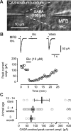
Figure 1.
Figure 1.
GABA-evoked currents in hippocampal mossy fibers. A, The stimulation and recording (rec.) configuration. Direct presynaptic recordings from MFBs at 34°C and focal puff application of GABA through a second glass pipette are shown. B, Top, Average traces of GABA-evoked presynaptic inward currents (symmetrical chloride condition) and bath application of the GABAA receptor antagonist bicuculline (Bic). Ticks indicate puff application. Bottom, Peak current amplitude plotted over recording time. C, GABA-evoked peak current amplitudes in mossy fibers of animals aged between 12 d and 3 months. rec., Recording; ampl., amplitude.
Figure 2.
Figure 2.
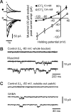
GABAA receptor-mediated currents are generated on the mossy fiber. A, Left, Muscimol-evoked currents recorded with a low (top trace) and a high (bottom trace) intracellular chloride concentration at different presynaptic holding potentials. Right, Muscimol-evoked currents plotted over the presynaptic holding potential for the low (filled symbols) and the high (open symbols) intracellular chloride condition (each n = 5). ampl., Amplitude. B, MFB recording in the whole-cell configuration before and during focal application of muscimol to the MFB via a second pipette. C, Recording from an outside-out patch of an MFB before and during bath application of GABA.
Figure 3.
Figure 3.
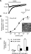
Basic characterization of presynaptic GABAA receptors on mossy fibers. A, Focal application of GABA for 3 s at two different GABA concentrations. B, Dose–response curve for GABA applied to presynaptic GABAA receptors. The peak current amplitude of puff-evoked currents normalized to that evoked with 10 mm GABA (Ref.) is plotted over the respective GABA “test” concentration (Test; n = 3 for each test concentration). Test and Ref. were applied interleaved by two pipettes (see inset). C, The benzodiazepine binding site agonist zolpidem was bath applied (bath-appl.) at increasing concentrations during GABA puff application with 10 and 100 μm GABA (each n = 4).
Figure 4.
Figure 4.
Tonic activation of presynaptic GABAA receptors is prevented by GAT 1. A, Mossy fiber input resistance was monitored using a long hyperpolarizing current pulse (top) and recording the resulting voltage traces (bottom) during perfusion with ACSF (baseline condition; black traces) and with either 10 μm bicuculline (BIC; middle, gray trace) or 1 μm NNC 711 (bottom, gray trace) added to the ACSF. B, Bath application of the GAT 1 blocker (NNC 711, 1 μm) during puff application of GABA to an MFB. Current traces are shown superimposed.
Figure 5.
Figure 5.
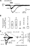
High-frequency stimulation in CA3-SR elicits transient GABAA receptor-mediated spill-over currents in MFBs. A, Twenty pulses at 100 Hz applied to the CA3-SR. Gray traces, Successive bath application of the GAT 1 blocker NNC 711 (1 μm) and the GABAA receptor antagonist gabazine (10 μm). rec., Recording. B, Bar graph summarizing the effect of bath application of zolpidem (ZPD; 2 μm; n = 2), NNC 711 (1 μm; n = 5), and gabazine (3–10 μm; n = 7) to the peak current amplitude of spill-over currents and the effect of lowered stimulation frequency (30 Hz) in ACSF and NNC 711 (n = 3). rel., Relative, basel., baseline. C, Left, Spill-over currents recorded at different presynaptic holding potentials (U h). Right, Normalized (Norm.) spill-over peak current amplitude (open symbols) plotted over the presynaptic holding potential. The filled symbol indicates the effect of gabazine application (10 μm). stim., Stimulation; ampl., amplitude.
Figure 6.
Figure 6.
Presynaptic GABA spill-over is sensitive to CB1 receptor activation. A, Averaged spill-over currents recorded at an MFB. Pharmacological conditions are indicated above the traces. stim., Stimulation. B, Spill-over peak current amplitude (ampl.) is plotted over the recording time. Horizontal bars indicate application of CB1 receptor agonist WIN 55,212-2 (2 μm), CB1 receptor antagonist AM 251 (10 μm), and gabazine (10 μm). C, Summary bar graph of experiments shown in B (n = 4) and of experiments in which the group 2 metabotropic glutamate receptor agonist DCG-4 (1 μm) was bath applied (n = 4). Experiments were done in the presence of 2 μm zolpidem and 1 μm NNC 711. WIN, WIN 55,212-2; AM, AM 251; GZ, gabazine.
Figure 7.
Figure 7.
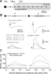
Simulations of activating presynaptic GABAA receptors on single and multiple MFBs including axonal segments. A, Scheme illustrating the spatial relationship of mossy fibers (pearl chain) and the axonal arborization (gray shaded area) of mossy fiber-associated interneurons (IN) (Vida and Frotscher, 2000) in CA3 stratum lucidum (CA3-SL). GCL, Granule cell layer. B, Top, Scheme indicating activation of presynaptic GABAA receptors (gray box) on a single MFB including 75 μm of mossy fiber in both directions (left) or on four MFBs including corresponding axonal segments (right). Middle, Corresponding simulated presynaptic GABAA receptor-mediated conductance (gGABAA) changes. Bottom, GABA-EPreSPs. The asterisks indicate the MFB from which GABA-EPreSP is shown. Three different reversal potentials of the GABAA receptor-mediated current were assumed (see inset). U rev, Reversal potential; U 0, resting potential. C, Corresponding maximal axonal membrane depolarizations (at ∼50 ms after the onset of gGABA) plotted over the axonal distance from the granule cell soma. The line code corresponds to the inset in B. Membr. pot., Membrane potential.

References

    1. Alger BE, Nicoll RA. GABA-mediated biphasic inhibitory responses in hippocampus. Nature. 1979;281:315–317. - PubMed
    1. Alle H, Geiger JRP. Combined analog and action potential coding in hippocampal mossy fibers. Science. 2006;311:1290–1293. - PubMed
    1. Awatramani GB, Price GD, Trussell LO. Modulation of transmitter release by presynaptic resting potential and background calcium levels. Neuron. 2005;48:109–121. - PubMed
    1. Banks MI, Pearce RA. Kinetic differences between synaptic and extrasynaptic GABAA receptors in CA1 pyramidal cells. J Neurosci. 2000;20:937–948. - PMC - PubMed
    1. Berger T, Schwarz C, Kraushaar U, Monyer H. Dentate gyrus basket cell GABAA receptors are blocked by Zn2+ via changes of their desensitization kinetics: an in situ patch-clamp and single-cell PCR study. J Neurosci. 1998;18:2437–2446. - PMC - PubMed

Publication types

MeSH terms

LinkOut - more resources