. 2007 Feb 20;46(7):1821-8.
doi: 10.1021/bi6024403. Epub 2007 Jan 25.

Structural and functional analysis of the pyocyanin biosynthetic protein PhzM from Pseudomonas aeruginosa

Affiliations

Structural and functional analysis of the pyocyanin biosynthetic protein PhzM from Pseudomonas aeruginosa

James F Parsons et al. Biochemistry. .

Abstract

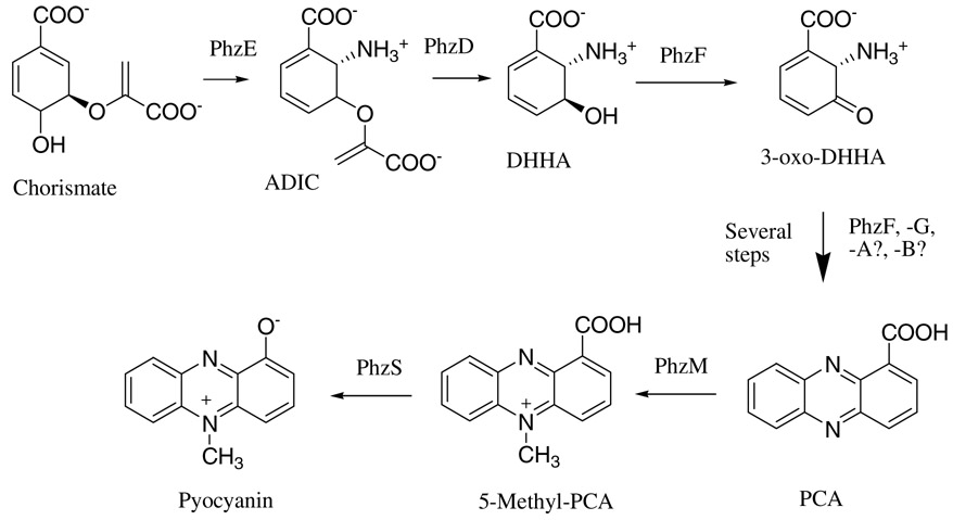
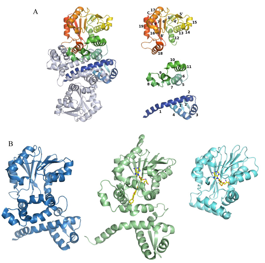
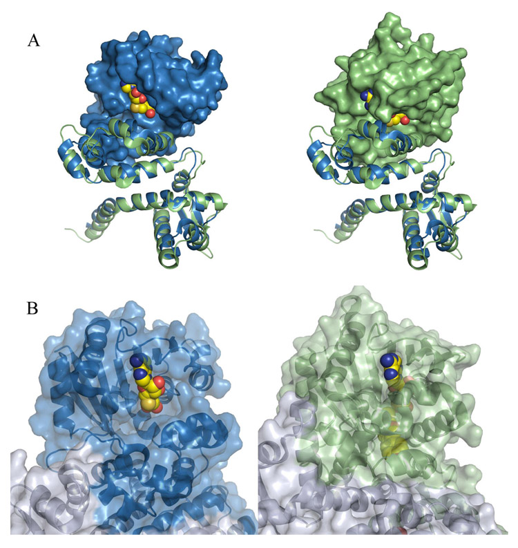
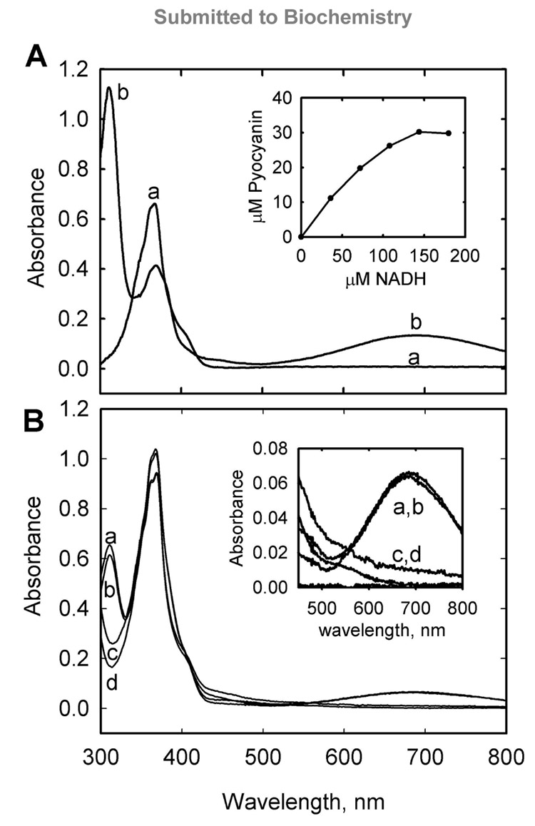
Pyocyanin is a biologically active phenazine produced by the human pathogen Pseudomonas aeruginosa. It is thought to endow P. aeruginosa with a competitive growth advantage in colonized tissue and is also thought to be a virulence factor in diseases such as cystic fibrosis and AIDS where patients are commonly infected by pathogenic Pseudomonads due to their immunocompromised state. Pyocyanin is also a chemically interesting compound due to its unusual oxidation-reduction activity. Phenazine-1-carboxylic acid, the precursor to the bioactive phenazines, is synthesized from chorismic acid by enzymes encoded in a seven-gene cistron in P. aeruginosa and in other Pseudomonads. Phenzine-1-carboxylic acid is believed to be converted to pyocyanin by the sequential actions of the putative S-adenosylmethionine-dependent N-methyltransferase PhzM and the putative flavin-dependent hydroxylase PhzS. Here we report the 1.8 A crystal structure of PhzM determined by single anomalous dispersion. Unlike many methyltransferases, PhzM is a dimer in solution. The 36 kDa PhzM polypeptide folds into three domains. The C-terminal domain exhibits the alpha/beta-hydrolase fold typical of small molecule methyltransferases. Two smaller N-terminal domains form much of the dimer interface. Structural alignments with known methyltransferases show that PhzM is most similar to the plant O-methyltransferases that are characterized by an unusual intertwined dimer interface. The structure of PhzM contains no ligands, and the active site is open and solvent-exposed when compared to structures of similar enzymes. In vitro experiments using purified PhzM alone demonstrate that it has little or no ability to methylate phenzine-1-carboxylic acid. However, when the putative hydroxylase PhzS is included, pyocyanin is readily produced. This observation suggests that a mechanism has evolved in P. aeruginosa that ensures efficient production of pyocyanin via the prevention of the formation and release of an unstable and potentially deleterious intermediate.


Figures

Figure 1
Figure 1
Proposed biosynthetic pathway leading to pyocyanin in P. aeruginosa.
Figure 2
Figure 2
Structure of the PhzM dimer, illustration of the secondary structural elements of PhzM, and a comparison of the PhzM monomer with representative methyltransferases. (A) The PhzM dimer, one subunit in rainbow coloration, the other in gray. Secondary structure diagram of the PhzM monomer separated by domain and colored as in the dimer. Helices are numbered 1–18, strands are numbered 1'–9'. (B) Ribbon diagrams comparing the structure of PhzM with those of a structural homolog (IOMT), and a typical monomeric small molecule methyltransferase, YecO, from Heamophilus influenzae.
Figure 3
Figure 3
Surface representations illustrating the solvent exposed state of the PhzM active site. (A) Superposition of PhzM and IOMT. In both panels, domains one and two were aligned and are shown as blue (PhzM) and green (IOMT) ribbons in order to indicate clearly that the viewing angle is identical in each panel. In the left panel the surface of domain three of PhzM only is shown along with SAH from IOMT. In the right panel, the surface of domain three of IOMT is shown along with bound SAH. (B) Portions of the PhzM structure (right) with a model of SAH in the active site and IOMT (left) in complex with products shown with transparent surfaces. Modeled or bound ligands are shown as space-filling spheres.
Figure 4
Figure 4
A coupled reaction of PhzM and PhzS converts phenazine-1-caboxylic acid to pyocyanin. Panel A shows spectra observed prior to the addition of NADH (a) and subsequent to titration with NADH (b). Typical absorption of PCA in pH 7.8 reaction buffer at 370 nm is observed prior to NADH addition while typical absorption of pyocyanin at 310 nm, 370 nm, and 690 nm (broad) is observed at the end of the reaction. Wavelengths below 300 nm are obscured by absorbance of the PhzM cofactor, S-adenosyl-methionine, and its reaction products. Inset is a plot of pyocyanin production plotted versus NADH consumption for the reaction indicating that more than stoichiometric amounts of NADH were required to approach total conversion of PCA in the reaction (34 µM) to pyocyanin. Panel B shows the absorption spectra of reactions carried out in equilibrium dialysis chambers separated by 8000 molecular weight cutoff dialysis membranes. Spectra (a) and (b) were obtained from solutions on separate sides of a chamber that initially had PhzM and PhzS on one side and PCA on the other. Spectra (c) and (d) were obtained from solutions on separate sides of a chamber that initially had PhzM and PhzS on opposite sides of the membrane and PCA on both sides. Inset is focused on the absorbance at 690 nm indicative of pyocyanin in samples (a) and (b). Complete conversion of PCA to pyocyanin was not observed in the experiments depicted in panel B due to pyocyanin and PhzS mediated oxygen and NADH depletion (see text).

References

    1. Laursen JB, Nielsen J. Phenazine Natural Products: Biosynthesis, Synthetic Analogues, and Biological Activity. Chem. Rev. 2004;104:1663–1686. - PubMed
    1. Lau GW, Ran H, Kong F, Hassett DJ, Mavrodi D. Pseudomonas aeruginosa pyocyanin is critical for lung infection in mice. Infect. Immun. 2004;72:4275–4278. - PMC - PubMed
    1. Van Delden C, Iglewski BH. Cell-to-cell signaling and Pseudomonas aeruginosa infections. Emerg. Infect. Dis. 1998;4:551–560. - PMC - PubMed
    1. Zelenitsky SA, Harding GK, Sun S, Ubhi K, Ariano RE. Treatment and outcome of Pseudomonas aeruginosa bacteraemia: an antibiotic pharmacodynamic analysis. J. Antimicrob. Chemother. 2003;52:668–674. - PubMed
    1. Reszka KJ, Denning GM, Britigan BE. Photosensitized oxidation and inactivation of pyocyanin, a virulence factor of Pseudomonas aeruginosa. Photochem. Photobiol. 2006;82:466–473. - PubMed

Publication types

Associated data

LinkOut - more resources