. 2007 Jan 25:4:13.
doi: 10.1186/1743-422X-4-13.

Demonstration of infectious salmon anaemia virus (ISAV) endocytosis in erythrocytes of Atlantic salmon

Affiliations

Demonstration of infectious salmon anaemia virus (ISAV) endocytosis in erythrocytes of Atlantic salmon

Samuel T Workenhe et al. Virol J. .

Abstract

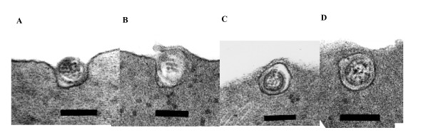
Infectious salmon anaemia (ISA) virus (ISAV) is a fish orthomyxovirus that has recently been assigned to the new genus Isavirus within the family Orthomyxoviridae. It possesses the major functional characteristics of the virus family including haemagglutinating, receptor destroying enzyme (RDE), and fusion activities associated with the virion surface proteins. It is generally accepted that ISAV agglutinates erythrocytes of several fish species and that the ISAV RDE activity dissolves this haemagglutination reaction except for Atlantic salmon (Salmo salar) erythrocytes. We used electron microscopy to examine the physical interaction between ISAV and erythrocytes from Atlantic salmon and rainbow trout (Oncorhynchus mykiss) during haemagglutination. We present evidence that ISAV enters into Atlantic salmon erythrocytes. Atlantic salmon erythrocytes incubated with ISAV for 4 hours showed endocytosis of the virus particles, which is consistent with virus infection. These observations suggest that the lack of dissolution of ISAV-induced haemagglutination of Atlantic salmon erythrocytes favours virus infection of the erythrocytes. Moreover, such a haemagglutination-infection phenotype is fundamentally different from haemagglutination by avian and mammalian orthomyxoviruses, and is indicative of a different pathogenesis for the fish orthomyxovirus.


Figures

Figure 1
Figure 1
Transmission electron micrographs reveal the various stages of apparent endocytosis of the NBISA01 virus in Atlantic salmon erythrocytes. (A) a virus particle within an invagination of the plasma membrane (bar = 139 nm); (B) partial closure of the pit containing a virus particle (bar = 139 nm); (C) and (D) virus particle with in a vesicle in the erythrocyte cytoplasm (bar = 139 nm).
Figure 2
Figure 2
Amplification, melting curve, and agarose gel electrophoresis of RT-PCR targeting a 220-bp product on ISAV segment 8 using total RNA from haemagglutination tests at different sampling points. (A) Real time RT-PCR amplification curves (water (Ct = 0) – no template negative control; 1% rbc rt (Ct = 0) – rainbow trout erythrocyte control; 1% rbc as(Ct = 0) – Atlantic salmon erythrocyte control; 0 hr rt (Ct = 28.6 ± 4.62) – NBISA01 haemagglutination reaction of 1% rainbow trout erythrocyte sampled at 0 hour; 0 hr as(Ct = 30.8 ± 0.70) – NBISA01 haemagglutination reaction of 1% Atlantic salmon erythrocyte sampled at 0 hour; 18 hr rt (sample not run in triplicate because of insufficient total RNA) – NBISA01 haemagglutination reaction of 1% rainbow trout erythrocyte sampled after 18-hour incubation; 18 hr as (Ct = 32.62 ± 1.10) – NBISA01 haemagglutination reaction of 1% Atlantic salmon erythrocyte sampled after 18-hour incubation; 36 hr rt (Ct = 30.27 ± 0.63) – NBISA01 haemagglutination reaction of 1% rainbow trout erythrocyte sampled after 36-hour incubation; 36 hr as (Ct = 27.59 ± 0.70) – NBISA01 haemagglutination reaction of 1% Atlantic salmon erythrocyte sampled after 36-hour incubation; NBISA01 (Ct = 28.46 ± 0.52) – virus positive control). (B) melting curve of the real time RT-PCR of the run in (A). (C) PCR products resolved on 1% agarose gel electrophoresis and visualized by ethidium bromide staining. The order of the lanes in the gel picture is the same as in the amplification curves (A).

Similar articles

Cited by

References

    1. Moneke E, Groman DB, Wright GM, Stryhn H, Johnson GR, Ikede BO, Kibenge FSB. Correlation of virus replication in tissues with histologic lesions in Atlantic salmon Experimentally Infected with Infectious Salmon Anemia Virus. Vet Pathol. 2005;42:338–349. doi: 10.1354/vp.42-3-338. - DOI - PubMed
    1. Kibenge FSB, Kibenge MJT, Groman D, McGeachy S. In vivo correlates of Infectious salmon anemia virus pathogenesis in fish. J Gen Virol. 2006;87:2645–2652. doi: 10.1099/vir.0.81719-0. - DOI - PubMed
    1. Eliasen TM, Froystad MK, Dannevig BH, Jankowska M, Brech A, Falk K, Romeren K, Gjoren T. Initial events in infectious salmon anemia infection: Evidence for the requirement of a low-pH step. J Virol. 74:218–227. - PMC - PubMed
    1. Mikalsen AB, Sindere H, Mjaaland S, Rimstad E. Expression, antigenicity and studies on cell receptor binding of hemagglutinin of infectious salmon anemia virus. Arch Virol. 2005;150:1621–1637. doi: 10.1007/s00705-005-0502-4. - DOI - PubMed
    1. Falk K, Asperhaug V, Vlasak R, Endresen C. Identification and characterization of viral structural proteins of infectious salmon anemia virus. J Virol. 2004;78:3063–3071. doi: 10.1128/JVI.78.6.3063-3071.2004. - DOI - PMC - PubMed

Publication types

LinkOut - more resources