. 2007 Apr;26(1):56-65.
doi: 10.1016/j.nbd.2006.11.013. Epub 2007 Jan 23.

Aging-related changes in the nigrostriatal dopamine system and the response to MPTP in nonhuman primates: diminished compensatory mechanisms as a prelude to parkinsonism

Affiliations

Aging-related changes in the nigrostriatal dopamine system and the response to MPTP in nonhuman primates: diminished compensatory mechanisms as a prelude to parkinsonism

Timothy J Collier et al. Neurobiol Dis. 2007 Apr.

Abstract

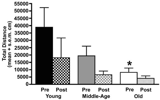
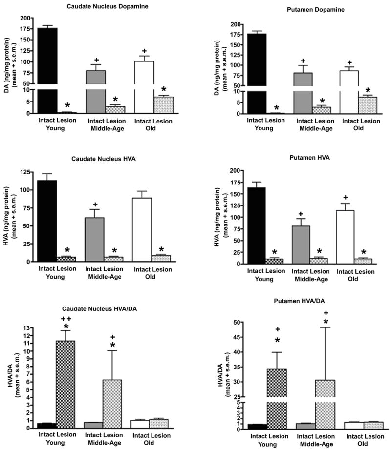
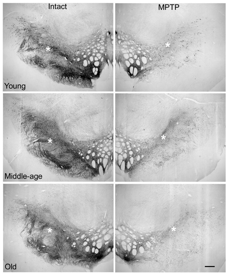
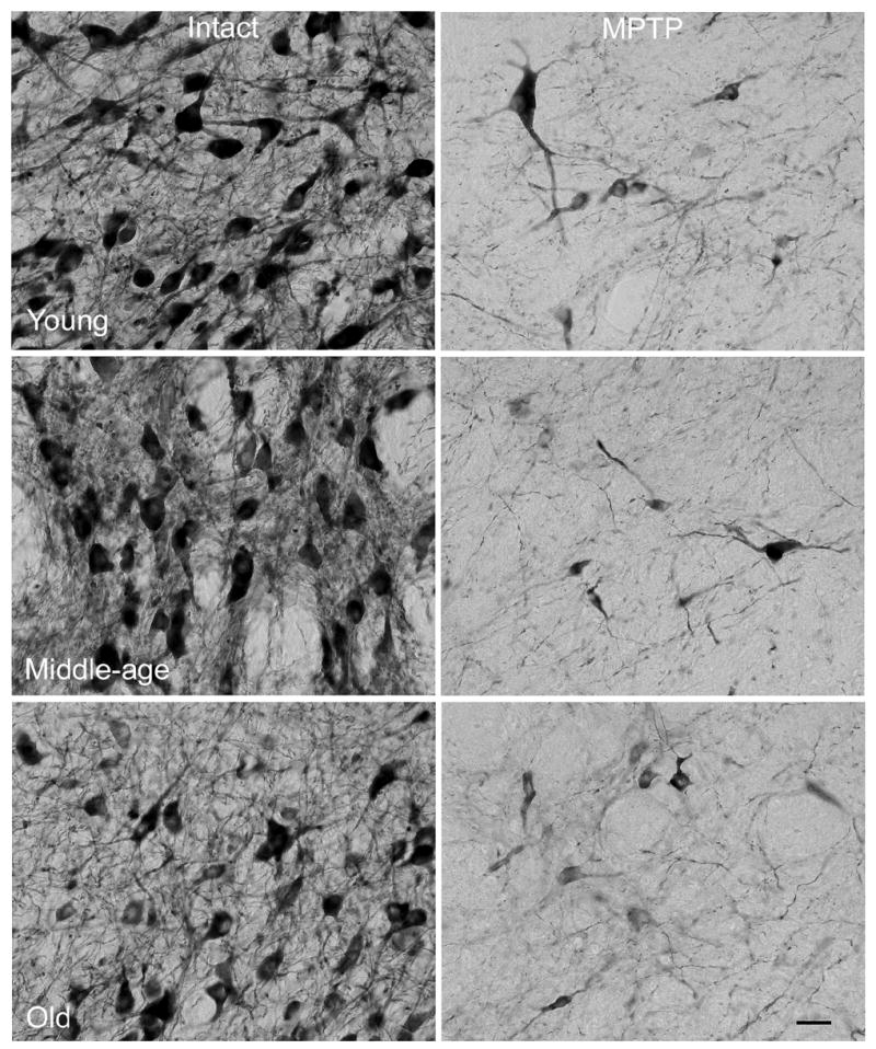
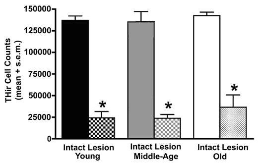
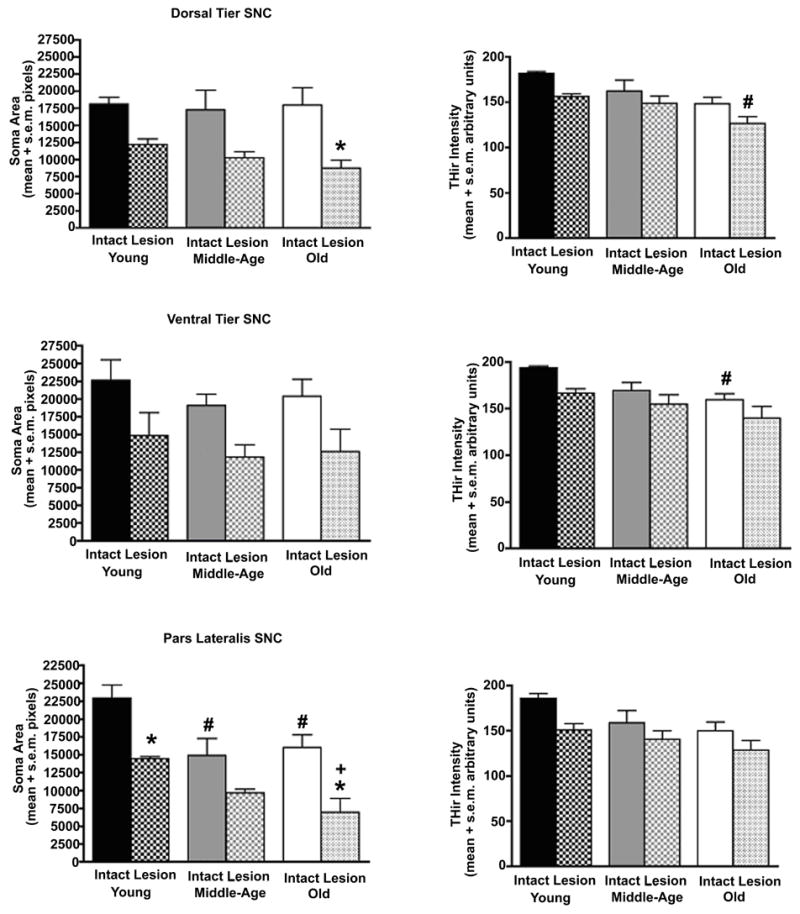
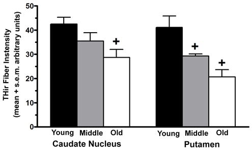
Aging is the most prominent risk factor for Parkinson's disease. Yet, consensus of how advancing age may predispose the dopamine (DA) system to parkinsonism is lacking. Three age ranges of female rhesus monkeys, 8-9, 15-17, and 21-31 years, received unilateral DA depletion with intracarotid 1-methyl-4-phenyl-1,2,3,6-tetrahydropyridine (MPTP). Morphological and biochemical analyses of DA-depleted and intact hemispheres revealed three primary findings: (1) The intact striatum exhibited age-related declines in dopamine (DA) and homovanillic acid (HVA) that were present by middle age; (2) In the MPTP-treated striatum, the compensatory increase in DA activity was absent in old monkeys; and (3) Age-associated morphological changes included declines in the density of tyrosine hydroxylase (TH) positive fibers in striatum, decreased nigral soma size, and optical density of TH, but no significant loss of neurons. These findings suggest that aging produces changes in the nigrostriatal DA system that approach the threshold for expression of parkinsonian features, and that progressive impairment of plasticity may be central to the role of aging in development of parkinsonism.


Figures

Figure 1
Figure 1
Locations of tissue punches for HPLC and regions analyzed for THir fiber staining. Tissue punches, 1.3mm in diameter (*), were taken from the dorsal lateral caudate nucleus and putamen at precommissural and commissural levels of the striatum for analysis of DA and HVA. A single 50X magnification microscopic field (rectangle) in caudate and putamen was analyzed for intensity of THir fiber staining in four coronal tissue sections from each subject. Tissue punches in ventral medial caudate and putamen were analyzed for striatal trophic factor activity reported in Collier et. al., 2005. Abbreviations: Cd = Caudate nucleus, IC = internal capsule, Pt = putamen.
Figure 2
Figure 2
Spontaneous locomotor activity progressively decreases with advancing age. Data represents total distance traveled (cm) in a video recording cage over a thirty minute interval prior to MPTP treatment (Pre) and 3 months after induction of unilateral parkinsonism (Post). Prior to MPTP treatment, spontaneous activity was noted to decrease progressively with advancing age, reaching statistical significance for the comparison of young adult animals to aged animals (* p=0.005). Further decreases in motor activity were noted after MPTP treatment, but these were not statistically different between age groups.
Figure 3
Figure 3
With advancing age, striatal DA content declines and the capacity to increase DA activity in response to insult is lost in the oldest monkeys. Significant aging-related decreases in DA content of the caudate nucleus and putamen of the intact hemisphere were detected in middle-age and sustained in old age. As expected, dramatic loss of striatal DA accompanied MPTP exposure in the opposite hemisphere (caudate and putamen: + p<0.0001 for comparison of young intact to middle-aged and aged intact; * p<0.0001 for comparison of lesioned hemisphere to intact hemisphere). Decreases in striatal HVA also accompanied aging and exposure to MPTP (caudate and putamen: + p<0.05 for comparison of young intact to middle-aged and aged intact; * p<0.0001 for comparison of lesioned hemisphere to intact hemisphere). Large increases in striatal DA activity followed exposure to MPTP in young and middle-aged monkeys, but this compensatory response was completely absent in aged monkeys (Caudate: * p<0.005 for comparison of intact to lesioned hemispheres, + p<0.009 for comparison of middle-age lesion to old lesion, ++ p<0.04 for comparison of young lesion to both middle-age and old lesion. Putamen: * p<0.005 for comparison of intact to lesioned hemispheres, + p<0.003 for comparison of young and middle-age lesion to old lesion).
Figure 4
Figure 4
Low magnification views of TH immunoreactivity in SNC of the intact hemisphere suggest a decrease in cell number and neuropil density accompany advancing age (calibration bar=600μm). However, quantitation of THir cell numbers with stereology indicates no aging-related loss of neurons. The difference in appearance at low magnification was accounted for by a progressive increase in the frequency of THir neurons that stained lightly and decreased in cross-sectional area with advancing age (Figure 5). The expected dramatic loss of THir SNC neurons accompanied MPTP exposure in the ipsilateral hemisphere. * denote regions associated with high magnification views presented in Figure 5.
Figure 5
Figure 5
With advancing age, the number of THir SNC neurons that stain lightly and exhibit lower cross-sectional area increase in the intact hemisphere. While THir intensity and soma area are preserved through middle-age, old subjects exhibited increased variability in these measures. SNC neurons surviving the MPTP insult in the ipsilateral hemisphere exhibited variable further declines in THir intensity and soma area. * in Figure 4 depict locations of higher magnification views presented here. Calibration bar = 50μm.
Figure 6
Figure 6
Counts of THir neurons using stereology reveal no aging-related change in the intact hemisphere of monkeys. The expected significant loss of THir neurons accompanied exposure to MPTP. The magnitude of this loss was not different among age groups (* p<0.0001 for comparison of intact to lesioned hemisphere).
Figure 7
Figure 7
Aging and exposure to MPTP produce specific reductions in soma area and TH staining intensity in the three major subdivisions of SNC. For dorsal tier and ventral tier neurons of the intact hemisphere, there was an aging-related reduction in intensity of THir that reached statistical significance for the comparison of young adult monkeys to aged monkeys (dorsal tier and ventral tier: # p<0.02 for comparison of young to old). For pars lateralis neurons an aging-related decline in THir was evident, but did not attain statistical significance (p=0.053). For these cells the change expressed was decreased soma cross-sectional area. This decline was detectable in middle-age and sustained in old age (# p<0.03). In general MPTP treatment was associated with further decreases in both THir intensity and soma area in all regions in all age groups. Statistically significant decreases in soma area were detected for dorsal tier neurons of aged monkeys (* p<0.01) and pars lateralis neurons of young and old monkeys (* p<0.03). This cell shrinkage after MPTP exposure was exaggerated in the oldest animals when compared to young adult animals (+ p<0.04).
Figure 8
Figure 8
The intensity of THir fiber staining decreased in caudate nucleus and putamen with advancing age in the intact hemisphere. Decreased fiber staining was more pronounced and expressed earlier in the putamen (+ Caudate: p<0.01 for comparison of young to old; Putamen: p<0.04 for comparison of young to middle-aged and old).

References

    1. Andersen AH, Zhang Z, Zhang M, Gash DM, Avison MJ. Age-associated changes in rhesus CNS composition identified by MRI. Brain Res. 1999;829:90–98. - PubMed
    1. Bankiewicz KS, Oldfield EH, Chiueh CC, Doppman JL, Jacobowitz DM, Kopin IJ. Hemiparkinsonism in monkeys after unilateral internal carotid artery infusion of 1-methyl-4-phenyl-1,2,3,6-tetrahydropyridine (MPTP) Life Sci. 1986;39:7–16. - PubMed
    1. Calne DB, Langston JW. Aetiology of Parkinson’s disease. Lancet. 1983;2:1457–1459. - PubMed
    1. Carvey PM, Punati A, Newman MB. Cell Transplant. Vol. 15. 2006. Progressive dopamine neuron loss in Parkinson’s disease: The multiple hit hypothesis; pp. 239–250. - PubMed
    1. Collier TJ, Ling ZD, Carvey PM, Fletcher-Turner A, Yurek DM, Sladek JR, Jr, Kordower JH. Striatal trophic factor activity in aging monkeys with unilateral MPTP-induced parkinsonism. Exper Neurol. 2005;191:S60–S67. - PubMed

Publication types

MeSH terms