. 2007 Jan 26;128(2):309-23.
doi: 10.1016/j.cell.2006.12.029.

FoxOs are lineage-restricted redundant tumor suppressors and regulate endothelial cell homeostasis

Affiliations

FoxOs are lineage-restricted redundant tumor suppressors and regulate endothelial cell homeostasis

Ji-Hye Paik et al. Cell. .

Abstract

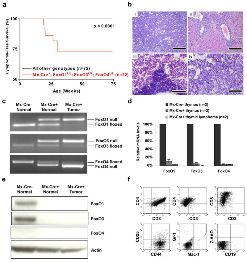
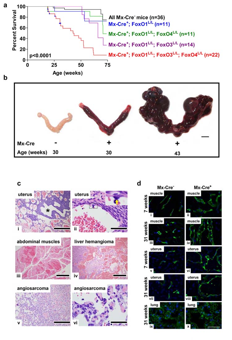
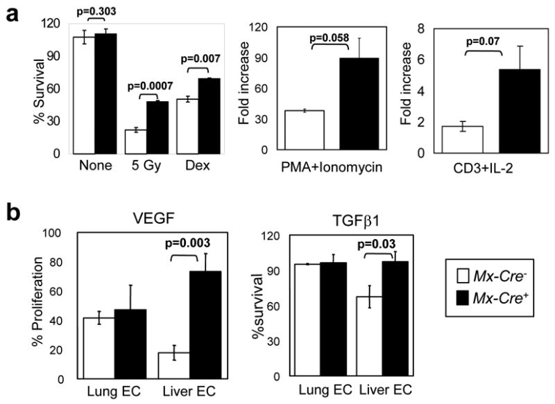
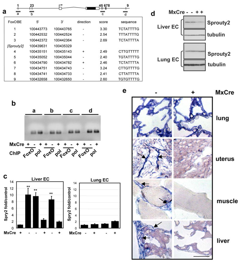
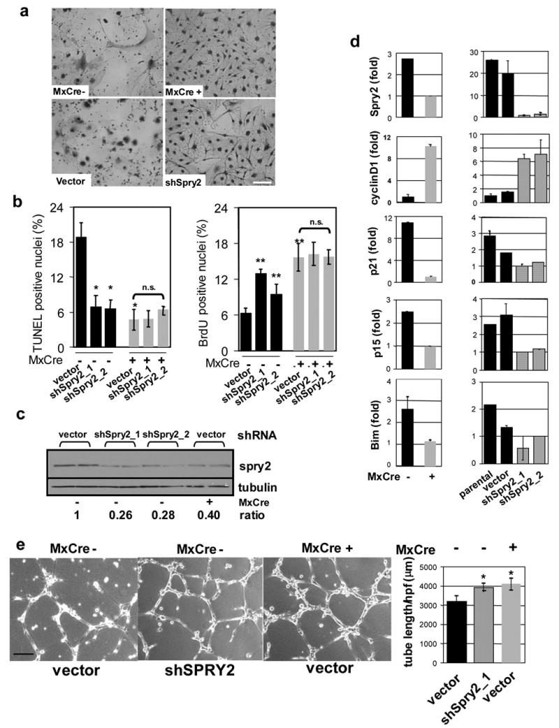
Activated phosphoinositide 3-kinase (PI3K)-AKT signaling appears to be an obligate event in the development of cancer. The highly related members of the mammalian FoxO transcription factor family, FoxO1, FoxO3, and FoxO4, represent one of several effector arms of PI3K-AKT signaling, prompting genetic analysis of the role of FoxOs in the neoplastic phenotypes linked to PI3K-AKT activation. While germline or somatic deletion of up to five FoxO alleles produced remarkably modest neoplastic phenotypes, broad somatic deletion of all FoxOs engendered a progressive cancer-prone condition characterized by thymic lymphomas and hemangiomas, demonstrating that the mammalian FoxOs are indeed bona fide tumor suppressors. Transcriptome and promoter analyses of differentially affected endothelium identified direct FoxO targets and revealed that FoxO regulation of these targets in vivo is highly context-specific, even in the same cell type. Functional studies validated Sprouty2 and PBX1, among others, as FoxO-regulated mediators of endothelial cell morphogenesis and vascular homeostasis.


Figures

Figure 1
Figure 1. Thymic lymphomas in mice following somatic deletion of three FoxO genes
a, Thymic lymphoma-free survival of pI-pC treated Mx-Cre+ mice and controls representing combined genotypes including: Mx-Cre+;FoxO1L/L (n=11), Mx-Cre+;FoxO1/3L/L (n=14), Mx-Cre+;FoxO1/4L/L (n=11), and all Mx-Cre controls (n=36). No lymphomas were observed in controls up to 100 weeks of age. b, Histology and tissue infiltration of thymic lymphoma in Mx-Cre+ mouse, H&E stains. (i) thymus (ii) liver, (iii) lung, and (iv) bone marrow. Scale bars: 200 μm (i and ii) and 100 μm (iii and iv). c, FoxO1, FoxO3, and FoxO4 gene deletions in thymic lymphomas and control thymi by PCR analysis. d, Reduction of mRNA levels of FoxO1, FoxO3, and FoxO4 in Mx-Cre+ endothelial cells. Quantitative real-time PCR performed on Mx-Cre thymi (7 weeks), Mx-Cre+ thymi (7 weeks), and Mx-Cre+ thymic lymphomas (n=2, 19 and 30 weeks post pI-pC); relative reduction of FoxO levels relative to Mx-Cre thymi is shown. e, Western blot analysis of Mx-Cre and Mx-Cre+ thymi and thymic lymphoma samples. f, Flow cytometric analysis of representative thymic lymphoma.
Figure 2
Figure 2. Systemic hemangiomas in mice following somatic deletion of three FoxO alleles
a, Survival of Mx-Cre+ and Mx-Cre littermates controls. Blue squares indicate deaths due to thymic lymphomas. P value indicates comparison between Mx-Cre+ mice (red) and all Mx-Cre controls (black). b, Age-dependent progression of uterine vascular lesions in Mx-Cre+ females, scale Bar=5 mm. c, Histology of systemic vascular lesions, H&E stains. Genotypes are Mx-Cre+ unless otherwise noted. i, uterus, low magnification (asterisk: uterine lumen); ii, uterine hemangioma (inset: benign endothelium of hemangioma vascular channels; arrow: endothelial cell); iii, abdominal muscle hemangioma; iv, liver hemangioma, Mx-Cre+ mouse; v-vi, angiosarcoma, Mx-Cre+ mouse. Scale bars: 500 μm (i and v), 400 μm (iv), 200 μm (iii), and 50 μm (ii and vi). d, Fluorescence micrographs of abdominal muscle, uterine horn, and lung endothelium after vascular perfusion of fluorescein-labeled lectin (blue: DAPI labelled nuclei). Note increased endothelial cell density by 7 weeks in muscle and uterine horn but not in lung as late as 31 weeks. Scale bar=50 μm.
Figure 3
Figure 3. Functional studies of thymocytes and endothelial cells rendered null for all three FoxOs
a, Increased proliferation and defective induction of apoptosis in Mx-Cre+ thymocytes. Thymocytes from 8-week old mice (n=3 per genotype) were plated and the indicated mitogens or apoptotic stimuli were applied. Experiment was performed twice with similar results; data from a representative experiment is shown. Survival % was calculated after 18hrs of treatment (5 Gy: γ-irradiation; Dex: 1 μM Dexamethasone). b, Analysis of viability of endothelial cells. MTT assays were performed on Mx-Cre and Mx-Cre+ lung (n=2) and liver (n=3) ECs. Y axis, normalized response (%) per OD595. Results represent two independent experiments. For a and b, data are mean ± SEM.
Figure 4
Figure 4. Regulation of Sprouty2 expression in liver ECs by FoxOs
a, Multiple conserved FoxO binding sites (predicted) in vicinity of Sprouty2 gene; ~11kb of the genomic region is shown. 5 of 10 sites were conserved in >3 species including human and mouse (supplementary methods). Sites were scored by 10-based logarithm of the likelihood ratio between the FoxO and background models. b. The FoxO BE occupancy by FoxOs. Soluble chromatin was prepared from Mx-Cre and Mx- Cre+ liver ECs and immunoprecipitated with a mixture of antibodies against the 3 FoxOs. Anti-pol II and anti-FoxO immunoprecipitated DNA (FoxO) was amplified giving rise to ~110bp products (a-d, illustrated in panel a). c, Quantitative real-time PCR analysis for Sprouty2. Expression of Sprouty2 is tightly correlated with FoxO deletion (Mx-Cre+) in liver ECs but not in lung ECs (**, p<0.01); error bars = s.e. d. Immunoblot analysis for Sprouty2. Expression of Sprouty2 is lower in FoxO deletion (Mx-Cre+) in liver ECs but not in lung ECs compared with control ECs. Duplicate loading shown for each EC line. e. RNA in situ hybridization using antisense Sprouty2 probe tissues from Mx-Cre and Mx-Cre+ mice 3 weeks after pI-pC injection. Arrows point to EC with strong Sprouty2 expression (bar=20μm).
Figure 5
Figure 5. FoxOs regulate liver EC angiogenic response through Sprouty2
a, Growth advantage of Mx-Cre+over Mx-Cre liver ECs after 10 days culture. Sprouty2 knockdown in Mx-Cre liver ECs had similar effect. Bar=15μm. b, TUNEL and BrdU. Knockdown of Sprouty2 (shSpry2_1 &_2, shRNAs) in Mx-Cre liver ECs phenocopies cell growth and apoptosis phenotypes in Mx-Cre+ liver ECs; * indicates p<0.01; error bars represent ± s.e. c, knock-down of endogenous Sprouty2 protein expression in Mx-Cre liver ECs by shSpry2. Two independent replicates are shown. Band densities were measured by ImageJ and normalized to tubulin. Ratio indicates normalized values over that of control (vector infected Mx-Cre EC). d, Correlation of Sprouty2 expression with cyclinD1, p21, p15, Bim level in liver ECs by quantitative PCR analysis. Knockdown of Sprouty2 with two different shRNAs (shSpry2_1 &_2) recapitulates above differences for cyclinD1, p21, p15, and Bim relative to parental untreated and vector-only controls (lower panels). Results shown are from triplicate experiments; error bars represent ± s.e. e, VEGF-induced tube formation in liver ECs. Bar=100 μm. Average tubule length/HPF (±s.d.) measured by ImageJ software in multiple microscopic fields was plotted (*, p<0.01 versus vector infected Mx-Cre EC).

Comment in

References

    1. Anderson MJ, Viars CS, Czekay S, Cavenee WK, Arden KC. Cloning and characterization of three human forkhead genes that comprise an FKHR-like gene subfamily. Genomics. 1998;47:187–199. - PubMed
    1. Benezra R. Role of Id proteins in embryonic and tumor angiogenesis. Trends Cardiovasc Med. 2001;11:237–241. - PubMed
    1. Biggs WH, 3rd, Cavenee WK, Arden KC. Identification and characterization of members of the FKHR (FOX O) subclass of winged-helix transcription factors in the mouse. Mamm Genome. 2001;12:416–425. - PubMed
    1. Bikfalvi A, Klein S, Pintucci G, Rifkin DB. Biological roles of fibroblast growth factor-2. Endocr Rev. 1997;18:26–45. - PubMed
    1. Brunet A, Bonni A, Zigmond MJ, Lin MZ, Juo P, Hu LS, Anderson MJ, Arden KC, Blenis J, Greenberg ME. Akt promotes cell survival by phosphorylating and inhibiting a Forkhead transcription factor. Cell. 1999;96:857–868. - PubMed

Publication types

MeSH terms