. 2007 Apr 1;580(Pt 1):285-300.
doi: 10.1113/jphysiol.2006.125336. Epub 2007 Jan 25.

Inhibitory input from slowly adapting lung stretch receptors to retrotrapezoid nucleus chemoreceptors

Affiliations

Inhibitory input from slowly adapting lung stretch receptors to retrotrapezoid nucleus chemoreceptors

Thiago S Moreira et al. J Physiol. .

Abstract

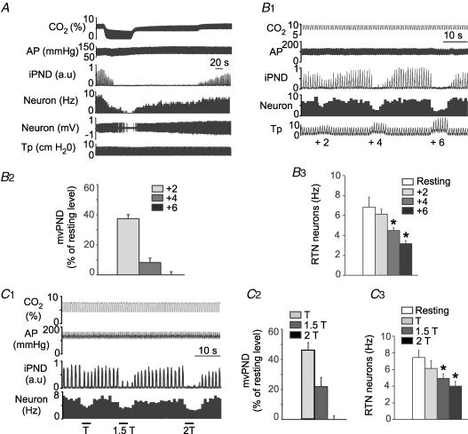
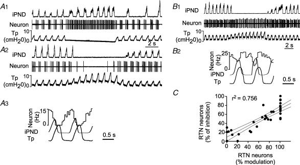
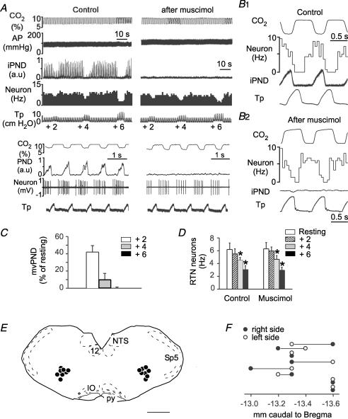
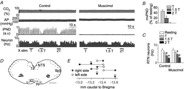
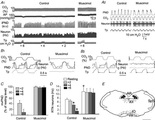
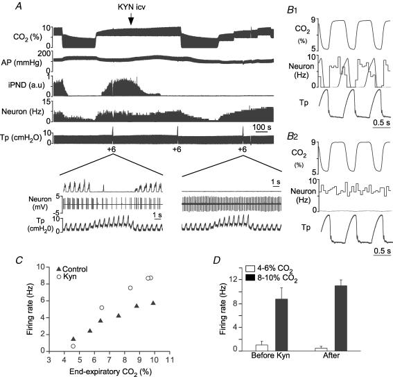
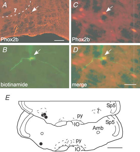
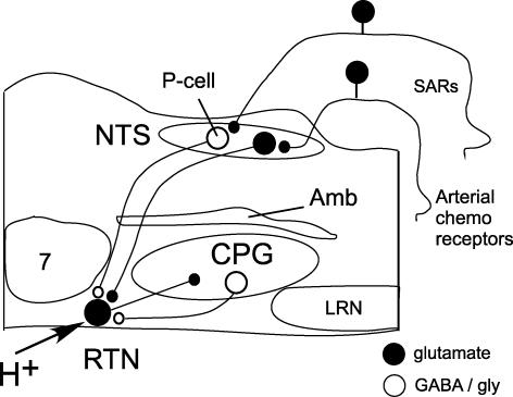
The retrotrapezoid nucleus (RTN) contains CO(2)-activated interneurons with properties consistent with central respiratory chemoreceptors. These neurons are glutamatergic and express the transcription factor Phox2b. Here we tested whether RTN neurons receive an input from slowly adapting pulmonary stretch receptors (SARs) in halothane-anaesthetized ventilated rats. In vagotomized rats, RTN neurons were inhibited to a variable extent by stimulating myelinated vagal afferents using the lowest intensity needed to inhibit the phrenic nerve discharge (PND). In rats with intact vagus nerves, RTN neurons were inhibited, also to a variable extent, by increasing positive end-expiratory pressure (PEEP; 2-6 cmH(2)O). The cells most sensitive to PEEP were inhibited during each lung inflation at rest and were instantly activated by stopping ventilation. Muscimol (GABA-A agonist) injection in or next to the solitary tract at area postrema level desynchronized PND from ventilation, eliminated the lung inflation-synchronous inhibition of RTN neurons and their steady inhibition by PEEP but did not change their CO(2) sensitivity. Muscimol injection into the rostral ventral respiratory group eliminated PND but did not change RTN neuron response to either lung inflation, PEEP increases, vagal stimulation or CO(2). Generalized glutamate receptor blockade with intracerebroventricular (i.c.v.) kynurenate eliminated PND and the response of RTN neurons to lung inflation but did not change their CO(2) sensitivity. PEEP-sensitive RTN neurons expressed Phox2b. In conclusion, RTN chemoreceptors receive an inhibitory input from myelinated lung stretch receptors, presumably SARs. The lung input to RTN may be di-synaptic with inhibitory pump cells as sole interneurons.


Figures

Figure 1
Figure 1. Inhibition of RTN CO2-sensitive neurons by lung inflation or low-intensity vagus nerve stimulation
A, example of one RTN neuron exposed to various levels of end-expiratory CO2 in a rat with intact vagus nerves. B1, effect of lung inflation (+2, +4 and +6 cmH2O positive end-expiratory pressure, PEEP) on the neuron shown in A. B2, effect of lung inflation (+2, +4 and +6 cmH2O) on neural minute × volume (mvPND) in a group of 28 rats with intact vagus nerves. B3, average effect of lung inflation (+2, +4 and +6 cmH2O) on the discharge rate of 36 RTN CO2-activated neurons sampled in these rats (*P < 0.05 relative to resting; RM ANOVA). C1, example of one RTN neuron recorded on the side ipsilateral to the stimulated vagal nerve in a vagotomized rat. This RTN neuron was detectably inhibited at the threshold (T) for PND inhibition. C2, group data showing the effect of vagus nerve stimulation (T, 1.5T and 2T) on mvPND (neural minute × volume) in 15 vagotomized rats. C3, group data showing the effect of vagus nerve stimulation (T, 1.5T and 2T) on the discharge rate of 18 RTN neurons in 15 vagotomized rats (one-way RM ANOVA: *P < 0.05 compared with resting). Abbreviations: AP, arterial pressure; iPND, integrated phrenic nerve discharge; Tp, tracheal pressure.
Figure 2
Figure 2. Respiratory pattern of RTN neurons inhibited by lung inflation
A, example of an RTN neuron that was strongly inhibited by increasing PEEP. A1, the neuron was instantly activated by interrupting the ventilator and became tonically active. A2, the neuron was 100% inhibited by lung inflation (+6 cmH2O PEEP). A3, peri-event histogram triggered on the tracheal pressure shows that the discharge probability of the neuron is zero at peak lung inflation. B, example of a different RTN neuron that was almost unaffected by increasing end-expiratory pressure. B1, a 6 cm increase in end-expiratory pressure produces little effect on this cell. B2, peri-event histogram triggered on the tracheal pressure showing absence of periodic slowing of the cell during lung inflation. C, correlation between the magnitude of the inhibition observed during each inflation cycle at rest (% modulation; abscissa) and the percentage reduction in mean firing rate caused by raising PEEP to 6 cmH2O (ordinate; 36 neurons). The first parameter (% modulation) was derived from peri-event-triggered histograms such as shown in A3. The value represents 1 minus the ratio between the discharge rate of the neuron at the peak of the tracheal pressure and the maximum discharge rate wherever it occurs during the respiratory cycle. For example the ratio was 1 (100%) in histogram A3 and 0 in histogram B2. The histograms were obtained at normal tidal volume and in the presence of 1 cm end-expiratory pressure. The 95% confidence intervals are also shown on the graph.
Figure 3
Figure 3. Bilateral injection of muscimol into the rostral ventral respiratory group (rVRG) does not change the effect of lung inflation on RTN neurons
A, this single RTN neuron was inhibited in a graded fashion by raising end-expiratory pressure to +2, +4 and +6 cmH2O. Bilateral injection of muscimol into the rVRG eliminated PND, raised arterial pressure (AP) but did not change markedly the inhibition of the RTN by lung inflation. Lower traces with expanded time scales indicate that muscimol did not change the phasic inhibition of the cell during lung inflation. B1, peri-event histogram of the discharge of the neuron shown in A. PND served as trigger. B2, peri-event histogram of the same RTN neuron after muscimol injection. In this case end-expiratory CO2 served as trigger. Note that the cell is still inhibited during the rise in tracheal pressure which corresponds to lung inflation. C, group data showing the effect of lung inflation (+2, +4 and +6 cmH2O PEEP) on mvPND before muscimol. D, group data showing the effect of lung inflation (+2, +4 and +6 cmH2O PEEP; 1 cm at rest) on the discharge rate of 8 RTN neurons before and after muscimol injection into the rVRG (2-way RM ANOVA; *P < 0.05 relative to resting level before or after muscimol; muscimol had no effect on the activity of the cells at rest or on the effect of PEEP). E, computer-assisted plots of the centre of the muscimol injection sites revealed by the presence of fluorescent microbeads included in the injectate. All sites projected on a single section (Bregma −13.3 mm). This level corresponds to the rVRG. Scale bar, 1 mm. F, rostro-caudal scatter of the muscimol injection into the ventrolateral medulla.
Figure 4
Figure 4. Bilateral injection of muscimol into the rostral ventral respiratory group (rVRG) does not change the inhibition of chemosensitive RTN neurons by low-intensity vagus nerve stimulation
A, a single RTN neuron was exposed to various levels of vagus nerve stimulation (T, threshold). Muscimol was then injected into the ventrolateral medulla on both sides causing a disappearance of PND. Muscimol had no effect on the resting discharge of the neuron and did not change its inhibition by vagus nerve stimulation. B, group data showing the effect of vagus stimulation on mvPND before muscimol (n = 8 rats). C, group data showing the effect of vagus stimulation on the discharge rate of the RTN neurons before and after muscimol injection into rVRG (n = 8 rats; 2-way RM ANOVA; *P < 0.05 relative to resting level before or after muscimol; muscimol had no effect on the activity of the cells at rest or on the effect of vagus nerve stimulation). D, computer-assisted plots of the centre of the muscimol injection sites revealed by the presence of fluorescent microbeads included in the injectate (coronal projection on plane Bregma −13.3 mm of the Paxinos atlas (Paxinos & Watson, 1998)). Scale bar, 1 mm. E, rostro-caudal scatter of muscimol injection into the ventrolateral medulla.
Figure 5
Figure 5. Bilateral injection of muscimol close to the tractus solitarius eliminates the effect of lung inflation on RTN neurons
A1, this RTN neuron was exposed to various levels of lung inflation (+2, +4 and +6 cmH2O). Muscimol injection next to the tractus solitarius on both sides raised the arterial pressure (AP) and eliminated the inhibition produced by lung inflation on PND and RTN neurons. A2, the RTN neuron had a clear pump rhythm before muscimol injection into the NTS. Muscimol injection into NTS dissociated the central respiratory generator from the ventilation cycle and eliminated the pump-related rhythm of the RTN neuron. B1, PND-triggered activity histogram of the RTN neuron shown in A, before and after muscimol was injected in the NTS. B2, tracheal pressure-triggered activity histogram of the same RTN neuron before and after muscimol injection. C, group data showing the effect of lung inflation (+2, +4 and +6 cmH2O PEEP) on mvPND before and after muscimol into the NTS. Lung inflation had no effect after muscimol (RM ANOVA). D, group data showing the effect of lung inflation (+2, +4 and +6 cmH2O) on the discharge rate of RTN neurons before and after muscimol into the NTS. The effect of lung inflation was significant before muscimol but not after (2-way RM ANOVA; *P < 0.05 relative to resting; +P < 0.05 relative to resting discharge before muscimol). E, computer-assisted plots of the centre of the muscimol injection sites revealed by the presence of fluorescent microbeads included in the injectate. All succesful injections were within or in close proximity of the solitary tract. Scale bar, 1 mm.
Figure 6
Figure 6. Kynurenate blocks the response to lung inflation but not the CO2 sensitivity of RTN neurons
A, example of one RTN neuron recorded before and after i.c.v. injection of 15 μmol of kynurenate (KYN). KYN eliminated PND, reduced BP, eliminated the neuron's response to lung inflation but had minimal effect on the cell's response to CO2. The expanded scale insets show the discharge pattern of the neuron before and after kynurenate. B1, peri-event histogram triggered on tracheal pressure demonstrating that, before kynurenate, the cell stops firing during each inflation of the lungs at rest (1 cmH2O PEEP). B2, similar histogram done after kynurenate showing that the cell is no longer inhibited by lung inflation. C, steady-state relationship between the activity of the cell shown in A and B and end-expiratory CO2 before and after KYN. D, resting discharge rate of 4 RTN neurons recorded at two levels of end-expiratory CO2 before and after i.c.v. kynurenate.
Figure 7
Figure 7. RTN neurons sensitive to lung inflation express Phox2b
A, cluster of Phox2b-ir nuclei that defines the RTN. The facial motor nucleus, 7, lacks detectable Phox2b immunoreactivity in adult rats. The ventral medullary surface is visible at the bottom. The y-shaped object at the lower left of the RTN is a blood vessel artifact. Scale bar, 100 μm. B, a single recorded cell labelled with biotinamide (arrow). C, Phox2b-ir nucleus of the labelled neuron (the arrow in A points to the same cell). D, merged image between B and C. Scale bar, 30 μm, applies to B–D. E, location of the five cells labelled juxtacellularly with biotinamide. Each cell had a Phox2b-ir nucleus. Scale bar, 1 mm. Other abbreviations: Amb, ambiguus nucleus; IO, inferior olive; py, pyramidal tract; Sp5, spinal trigeminal tract.
Figure 8
Figure 8. Presumed role of the RTN in chemosensory integration
RTN neurons are activated by CO2 via their intrinsic pH sensitivity and via inputs from the carotid bodies (Mulkey et al. 2004; Takakura et al. 2006). RTN neurons target various components of the CPG and are presumed to play a key role in breathing automaticity during anaesthesia and sleep. RTN in turn receives a feedback inhibition from the CPG represented here by a single inhibitory neuron located in the ventral respiratory column but known to consist of multiple types of cells (Guyenet et al. 2005a). Here, we propose that RTN neurons are inhibited by lung inflation via SAR activation and that the effect of SARs is mediated by GABAergic pump cells located close to the tractus solitarius at area postrema level. Abbreviations: Amb, nucleus ambiguus; CPG, respiratory pattern generator; LRN, lateral reticular nucleus; NTS, nucleus of the solitary tract; P-cell (pump cell), RTN, retrotrapezoid nucleus; SAR, slowly adapting pulmonary stretch receptors; 7, facial motor nucleus.

Comment in

  • Reflexively inhibiting respiratory drive.
    McCrimmon DR, Alheid GF. McCrimmon DR, et al. J Physiol. 2007 Apr 1;580(Pt 1):3. doi: 10.1113/jphysiol.2007.130500. Epub 2007 Feb 22. J Physiol. 2007. PMID: 17317738 Free PMC article. No abstract available.

Similar articles

Cited by

References

    1. Akilesh MR, Kamper M, Li A, Nattie EE. Effects of unilateral lesions of retrotrapezoid nucleus on breathing in awake rats. J Appl Physiol. 1997;82:469–479. - PubMed
    1. Bodineau L, Frugière A, Marlot D, Wallois F. Connections between retrotrapezoid nucleus and nucleus tractus solitarii in cat. Neurosci Lett. 2000a;280:111–114. - PubMed
    1. Bodineau L, Frugiere A, Marlot D, Wallois F. Effect of hypoxia on the activity of respiratory and non-respiratory modulated retrotrapezoid neurons of the cat. Auton Neurosci. 2000b;86:70–77. - PubMed
    1. Coleridge HM, Coleridge JC. Afferent innervation of lungs, airways, and pulmonary artery. In: Zucker IH, Gilmore JP, editors. Reflex Control of the Circulation. Boca Raton: CRC Press; 2001. pp. 579–607.
    1. Connelly CA, Ellenberger HH, Feldman JL. Respiratory activity in retrotrapezoid nucleus in cat. Am J Physiol. 1990;258:L33–L44. - PubMed

Publication types

MeSH terms