. 2007 Apr 1;580(Pt 1):133-47.
doi: 10.1113/jphysiol.2006.122739. Epub 2007 Jan 25.

Uncrossed actions of feline corticospinal tract neurones on lumbar interneurones evoked via ipsilaterally descending pathways

Affiliations

Uncrossed actions of feline corticospinal tract neurones on lumbar interneurones evoked via ipsilaterally descending pathways

E Jankowska et al. J Physiol. .

Abstract

Effects of stimulation of ipsilateral pyramidal tract (PT) fibres were analysed in interneurones in midlumbar segments of the cat spinal cord in search of interneurones mediating disynaptic actions of uncrossed PT fibres on hindlimb motoneurones. The sample included 44 intermediate zone and ventral horn interneurones, most with monosynaptic input from group I and/or group II muscle afferents and likely to be premotor interneurones. Monosynaptic EPSPs evoked by stimulation of the ipsilateral PT were found in 12 of the 44 (27%) interneurones, while disynaptic or trisynaptic EPSPs were evoked in more than 75%. Both appeared at latencies that were either longer or within the same range as those of disynaptic EPSPs and IPSPs evoked by PT stimuli in motoneurones, making it unlikely that premotor interneurones in pathways from group I and/or II afferents relay the earliest actions of uncrossed PT fibres on motoneurones. These interneurones might nevertheless contribute to PT actions at longer latencies. Uncrossed PT actions on interneurones were to a great extent relayed via reticulospinal neurones with axons in the ipsilateral medial longitudinal fascicle (MLF), as indicated by occlusion and mutual facilitation of actions evoked by PT and MLF stimulation. However, PT actions were also relayed by other supraspinal or spinal neurones, as some remained after MLF lesions. Mutual facilitation and occlusion of actions evoked from the ipsilateral and contralateral PTs lead to the conclusion that the same midlumbar interneurones in pathways from group I or II muscle afferents may relay uncrossed and crossed PT actions.


Figures

Figure 1
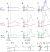
Figure 1. Examples of monosynaptic EPSPs evoked from the ipsilateral and contralateral pyramidal tracts (PTs)
Intracellular records from three interneurones and records from the cord dorsum (bottom traces). Averages of 10 single records. A and B show records from two interneurones in which EPSPs consistently followed successive ipsilateral PT stimuli. They are from a preparation with the medial longitudinal fascicle (MLF) intact. In the first neurone MLF stimuli evoked monosynaptic EPSPs (not illustrated) and in the second only longer latency PSPs shown in C. D, records from a preparation after transection of the MLF. Note that single and double stimuli applied to the contralateral PT evoked EPSPs at similarly long latencies and that only the 3rd stimulus evoked a disynaptic EPSP (at a shorter latency). Stimuli applied in the ipsilateral PT only evoked disynaptic EPSPs in this interneurone (at a latency of 4.93 ms; not illustrated). Dotted lines indicate onset of EPSPs. Square pulses at the beginning of the traces are calibration pulses, all of 0.2 mV. In this and the following figures the negativity is downward in intracellular records and upwards in cord dorsum records.
Figure 2
Figure 2. Comparison of latencies of EPSPs and IPSPs evoked from the ipsilateral PT, the contralateral PT and the MLF
A, comparison of latencies of EPSPs classified as evoked monosynaptically and disynaptically from the ipsilateral PT with latencies of EPSPs evoked monosynaptically from the MLF in the same neurones. The monosynaptic EPSPs of PT origin have been ranked in order of increasing latency. The data are for interneurones recorded in preparations with the MLF intact. Left ordinate, latencies from the 1st stimulus for the monosynaptic EPSPs and from the 2nd or 3rd stimulus for the disynaptic EPSPs. Right ordinate, equivalent conduction velocities of PT fibres evoking the monosynaptic EPSPs; their estimates were based on a conduction distance of 310 mm between the stimulation and recording sites, and 0.2 ms for utilization time (Jankowska & Roberts, 1972b) and 0.3 ms for one synaptic delay (Jankowska & Roberts, 1972a). For vertical lines see text. B, comparison of latencies of disynaptic IPSPs evoked from the ipsilateral PT (ranked in ascending order, the last two being probably evoked polysynaptically), the MLF and the contralateral PT, respectively. Data for 15 interneurones with the MLF intact and for 3 interneurones after transection of the MLF. In 14 of these interneurones monosynaptic and/or disynaptic EPSPs plotted in A and C preceded the IPSPs. C, comparison of latencies of disynaptic, trisynaptic and the last 1–3 most likely polysynaptic EPSPs from the ipsilateral PT (ranked in ascending order), the MLF and the contralateral PT, respectively. Data for 24 interneurones with the MLF intact (8 of which have been included in A) and 11 interneurones after transection of the MLF are plotted together because no statistically significant differences have been found between them. For vertical lines see text. The horizontal dotted lines in panels A, B and C indicate mean latency of monosynaptic EPSPs from the MLF.
Figure 3
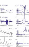
Figure 3. Disynaptic EPSPs from the ipsilateral PT and their occlusion with monosynaptic EPSPs from the MLF
Upper records are from an interneurone and lower records from the cord dorsum. Averages of 10 single records. A–C and D–F, effects of single, double and triple stimuli applied within the ipsilateral PT and ipsilateral MLF. Note that EPSPs from the MLF were evoked both monosynaptically and disynaptically (with the onset of disynaptic EPSPs at the level of the dotted line in E, showing the same latency as disynaptic EPSPs evoked by the 2nd PT stimulus in B). G–K, effects of PT and MLF stimuli, applied separately (G and I) and jointly (H), showing that the effects of the joint PT and MLF stimuli (H) were smaller than the sum of effects of these stimuli (J) when they were applied separately, with the difference in K. L–O, PSPs evoked from group I afferents (at 2T) and partly blocked spike potentials and PSPs from group II afferents (at 5T). Q, quadriceps; Sart, sartorius; PBST, posterior biceps–semitendinosus.
Figure 4
Figure 4. Disynaptic EPSPs evoked from the ipsilateral and contralateral PT and from the MLF
In each panel upper traces are intracellular records from an interneurone and the lower traces are records from the cord dorsum. Averages of 20 single records. AC, D–F and G–I, effects of single, double and triple stimuli applied within the ipsilateral PT, contralateral PT and ipsilateral MLF. Note that if any EPSPs followed the 1st stimuli, they were much smaller than those evoked by the 2nd and 3rd stimuli. Note also that EPSPs evoked by the 2nd and 3rd i PT, co PT and MLF stimuli were evoked at similar latencies (with onsets indicated by vertical dotted lines). J and K, examples of PSPs evoked from peripheral afferents. L and M, records from the same interneurone before it was penetrated, showing its synaptic but not antidromic activation by stimulation (1 mA) of either the ipsilateral (i) or the contralateral (co) lateral funiculus at the thoracic (Th) 13th level.
Figure 5
Figure 5. Mutual facilitation of disynaptic EPSPs evoked from the ipsilateral and contralateral PTs and from the MLF
In each panel upper traces are intracellular records from an interneurone (the same as in Fig. 4) and lower records from the cord dorsum. Averages of 20 single records. A–C, effects of stimuli applied to the ipsilateral PT or MLF alone and to both the PT and the MLF. D–F, as in A–C but for effects of the contralateral PT. Note hardly any effects of separate PT or MLF stimuli and potent facilitation of their effects when they were applied jointly. G, network of neurones likely to mediate both ipsilateral and contralateral actions of PT neurones on lumbar interneurones and motoneurones. Black circle represents premotor interneurones investigated in this study. The grey circle represents other spinal interneurones.
Figure 6
Figure 6. Examples of EPSPs from the ipsilateral and contralateral PT that were unlikely to be mediated by RS neurones
Averaged intracellular records from an interneurone and cord dorsum records. Note that the latency of the EPSP evoked from the ipsilateral PT that followed the 3rd stimulus (A) was longer than that of the EPSP evoked from the contralateral PT (B), and it was more than 3 ms longer than the latency of the monosynaptic EPSP from the MLF, and more than 2 ms longer than that of the disynaptic EPSP from the MLF (C).
Figure 7
Figure 7. Examples of IPSPs evoked by PT and MLF stimuli and mutual facilitation of effects of these stimuli
Intracellular records from two interneurones (A–I and J–L) and cord dorsum potentials (lower records in each panel). A–C, D–F, effects of decreasing numbers of stimuli applied to the ipsilateral PT and MLF. Vertical dotted lines indicate onset of IPSPs evoked by the 4th, 3rd and 2nd ipsilateral PT and the 3rd and 2nd MLF stimuli (with the indicated latencies). G, effects of stimulation of the contralateral PT. H and I, records showing short latency and low threshold input from group I afferents in the Q nerve. J–L, appearance of IPSPs when near-threshold ipsilateral PT stimuli preceded similarly near-threshold MLF stimuli which were ineffective when applied alone.
Figure 8
Figure 8. Mutual facilitation between effects of stimuli applied within the ipsilateral and the contralateral PT
Intracellular records from an interneurone and from the cord dorsum after transection of the MLF. Averages of 20 single records. A–D and E–H, effects of increasing numbers of stimuli, showing mixed excitatory–inhibitory effects from both PTs when at least 3 or 4 stimuli were applied and no effects of single and double stimuli. I, weak facilitation when one ipsilateral PT stimulus preceded two contralateral PT stimuli. J, stronger facilitation when two ipsilateral PT stimuli were combined with two contralateral PT stimuli. Dotted lines in I and J indicate latencies of the resulting EPSPs with respect to the first and second ipsilateral PT stimuli; note that they would be shorter with respect to co PT stimuli. K and L, monosynaptic EPSPs evoked by stimulation of the posterior biceps–semitendinosus (PBST) and plantaris (Pl) muscle afferents at 2T.
Figure 9
Figure 9. Examples of extracellular spikes and EPSPs evoked from the ipsilateral PT in a group Ia inhibitory interneurone in a preparation with the MLF intact
Top and middle traces, microelectrode records. Lower traces, records from the cord dorsum. A, extracellular field potential close to the interneurone; averaged records (n = 10). B, extracellular records from the interneurone; 5 traces superimposed. C, extracellular records from the interneurone; single record of responses to stimulation of the Q nerve at 400 Hz. D, monosynaptic EPSPs from Q after penetration of the neurone. E–H, intracellular records showing the effects of decreasing numbers of ipsilateral PT stimuli. Top, just after penetration of the interneurone. Middle, a few minutes after the penetration. Dotted vertical and horizontal lines indicate latencies of responses evoked by the indicated stimuli. Note the longer latency of EPSPs following the 2nd stimulus in F and G and shorter latencies of spikes (B) and EPSPs in E and F following the 3rd and 4th stimuli.
Figure 10
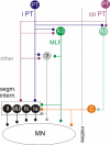
Figure 10. Diagram of neuronal organization in four so far investigated parallel pathways between PT neurones and ipsilateral motoneurones
These pathways are represented by disynaptic pathways via segmental interneurones (black neurones in the box), so far unidentified other neurones (grey neurone) and ipsilaterally descending RS neurones with axons in the MLF and by trisynaptic pathways via contralaterally descending RS neurones and commissural interneurones (labelled C; Edgley et al. 2004; Jankowska et al. 2006). Relay neurones in all these pathways appear to be used to mediate synaptic actions of both ipsilateral and contralateral PT neurones. For the sake of simplicity, somata of ipsilaterally and contralaterally descending PT and RS neurones are indicated to be located only on the left or the right sides, even though they are located on both sides. For further comments see text.

Similar articles

Cited by

References

    1. Alstermark B, Isa T, Tantisira B. Pyramidal excitation in long propriospinal neurones in the cervical segments of the cat. Exp Brain Res. 1991;84:569–582. - PubMed
    1. Alstermark B, Lundberg A, Pinter M, Sasaki S. Subpopulations and functions of long C3–C5 propriospinal neurones. Brain Res. 1987;404:395–400. - PubMed
    1. Brookhart JM, Morris RE. Antidromic potential recordings from the bulbar pyramid of the cat. J Neurophysiol. 1948;11:387–398. - PubMed
    1. Cabaj A, Stecina K, Jankowska E. Same spinal interneurons mediate reflex actions of group Ib and II afferents and crossed reticulospinal actions. J Neurophysiol. 2006;95:3911–3922. - PMC - PubMed
    1. Canedo A, Lamas JA. Pyramidal and corticospinal synaptic effects over reticulospinal neurones in the cat. J Physiol. 1993;463:475–489. - PMC - PubMed

Publication types

LinkOut - more resources