. 2007 Feb;170(2):427-35.
doi: 10.2353/ajpath.2007.060834.

Osteoclasts: what do they do and how do they do it?

Affiliations

Osteoclasts: what do they do and how do they do it?

Steven L Teitelbaum. Am J Pathol. 2007 Feb.

Abstract

As Americans live longer, degenerative skeletal diseases, such as osteoporosis, become increasingly prevalent. Regardless of cause, osteoporosis reflects a relative enhancement of osteoclast activity. Thus, this unique bone resorptive cell is a prominent therapeutic target. A number of key observations provide insights into the mechanisms by which precursors commit to the osteoclast phenotype and how the mature cell degrades bone. The osteoclast is a member of the monocyte/macrophage family that differentiates under the aegis of two critical cytokines, namely RANK ligand and M-CSF. Tumor necrosis factor (TNF)-alpha also promotes osteoclastogenesis, particularly in states of inflammatory osteolysis such as that attending rheumatoid arthritis. Once differentiated, the osteoclast forms an intimate relationship with the bone surface via the alphavbeta3 integrin, which transmits matrix-derived, cytoskeleton-organizing, signals. These integrin-transmitted signals include activation of the associated proteins, c-src, syk, Vav3, and Rho GTPases. The organized cytoskeleton generates an isolated microenvironment between the cell's plasma membrane and the bone surface in which matrix mineral is mobilized by the acidic milieu and organic matrix is degraded by the lysosomal protease, cathepsin K. This review focuses on these and other molecules that mediate osteoclast differentiation or function and thus serve as candidate anti-osteoporosis therapeutic targets.


Figures

Figure 1
Figure 1
First cure of patient with malignant osteopetrosis. The patient, a 3-month-old female, received a marrow transplant from her HLA/MLC compatible brother. Dramatic resolution of the sclerotic bone was evident within 7 weeks. The patient is well 27 years later. The presence of Y-chromosomes in her osteoclasts after transplant established the cell’s hematopoietic ontogeny in man (reprinted with permission from the N Engl J Med 1980, 302:701–708).
Figure 2
Figure 2
Osteoclast differentiation and function. Osteoblast lineage cells produce the osteoclastogenic cytokines RANKL and M-CSF, which recognize their respective receptors RANK and c-fms on macrophages, principally of marrow origin. OPG, also synthesized by osteoblast lineage cells, is a soluble decoy receptor that binds RANKL, thus preventing its interaction with RANK. RANKL and M-CSF are sufficient to promote the osteoclast phenotype. On contact with bone, the osteoclast polarizes via matrix-derived signals transmitted by the αvβ3 integrin, enabling the cell to form an isolated microenvironment between itself and the bone surface. The microenvironment is acidified by H+ATPase-mediated extracellular transport of protons. Intracellular pH is maintained by an electroneutral HCO3/Cl exchanger. The Cl entering the cell is released into the resorptive microenvironment by an ion channel charge coupled to the H+ATPase. The acidified microenvironment mobilizes the bone mineral, thereby exposing the organic phase of bone that is degraded by cathepsin K (modified and reprinted with permission from Science 2000, 289:1504–1508).
Figure 3
Figure 3
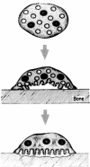
Formation of the osteoclast ruffled membrane. The unattached osteoclast contains numerous acidified vesicles bearing H+ATPases (proton pumps) illustrated as spikes. On attachment to bone, matrix-derived signals polarize the acidified vesicles to the bone-apposed plasma membrane into which they insert under the aegis of Rab3D. Insertion of the vesicles into the plasma membrane greatly increases its complexity and delivers the H+ATPases to the resorptive microenvironment.
Figure 4
Figure 4
αvβ3 integrin-deficient osteoclasts have an abnormal cytoskeleton. Both wild-type (β3+/+) and β3−/− osteoclasts contain tartrate-resistant acid phosphatase (red reaction product) and are multinucleated. Whereas wild-type osteoclasts spread in culture, those lacking αvβ3 fail to do so, manifesting a deranged cytoskeleton (reprinted with permission from the J Clin Invest 2000, 105:433–440).
Figure 5
Figure 5
Glucocorticoids disrupt the osteoclast cytoskeleton. Osteoclasts, generated on dentin in the presence and absence of dexamethasone (DEX), were stained with FITC-phalloidin to visualize the actin cytoskeleton. The well-demarcated actin rings present in naïve osteoclasts are disrupted by the glucocorticoid (reprinted with permission from the J Clin Invest 2006, 116:2152–2160).

References

    1. Whyte MP, Wenkert D, Clements KL, McAlister WH, Mumm S. Bisphosphonate-induced osteopetrosis. N Engl J Med. 2003;349:457–463. - PubMed
    1. Teitelbaum SL, Ross FP. Genetic regulation of osteoclast development and function. Nat Rev Genet. 2003;4:638–649. - PubMed
    1. Walker DG. Bone resorption restored in osteopetrotic mice by transplants of normal bone marrow and spleen cells. Science. 1975;190:784–785. - PubMed
    1. Walker DG. Spleen cells transmit osteopetrosis in mice. Science. 1975;190:785–787. - PubMed
    1. Coccia PF, Krivit W, Cervenka J, Clawson C, Kersey JH, Kim TH, Nesbit ME, Ramsay NK, Warkentin PI, Teitelbaum SL, Kahn AJ, Brown DM. Successful bone-marrow transplantation for infantile malignant osteopetrosis. N Engl J Med. 1980;302:701–708. - PubMed

Publication types

MeSH terms