. 2007 Feb;170(2):439-46.
doi: 10.2353/ajpath.2007.060219.

Protection against endotoxic shock as a consequence of reduced nitrosative stress in MLCK210-null mice

Affiliations

Protection against endotoxic shock as a consequence of reduced nitrosative stress in MLCK210-null mice

Hantamalala Ralay Ranaivo et al. Am J Pathol. 2007 Feb.

Abstract

This study investigated the consequences of deletion of the long isoform of myosin light chain kinase (MLCK210) on the cardiovascular changes induced by the bacterial endotoxin lipopolysaccharide (LPS) and cecal ligation puncture using MLCK210-/- mice. Here, we provide evidence that deletion of MLCK210 enhanced survival after intraperitoneal injection of LPS or cecal ligation puncture. LPS-induced vascular hyporeactivity to vasoconstrictor agents was completely prevented in aorta from MLCK210-/- mice. This was associated with a decreased up-regulation of nuclear facor-kappaB expression and activity, inducible nitric-oxide synthase, and level of oxidative stress in the vascular media. Furthermore, LPS-induced increase of nitric oxide production in the circulation and tissues (including heart, liver, and lung) that was correlated with an increased expression of inducible nitric-oxide synthase was also reduced in MLCK210-/- mice. These data demonstrate a role for MLCK210 in endotoxin shock injury associated with oxidative and nitrosative stresses and vascular hyporeactivity.


Figures

Figure 1
Figure 1
A: Survival curve of MLCK210+/+ and MLCK210−/− mice during 24 hours after LPS (40 mg/kg, i.p.) injection. **P < 0.01 was significant by log-rank test, n = 11 and 13 for MLCK210+/+ and MLCK210−/− mice, respectively. B: Survival curve of MLCK210+/+ and MLCK210−/− mice during 7 days after CLP ligation. *P < 0.05 was significant by log-rank test, n = 10 and 12 for MLCK210+/+ and MLCK210−/− mice, respectively. Changes in hemodynamic parameters: MAP (C) and heart rate (D) in response to LPS. There was no difference between both strains. Data represented mean ± SEM of six or seven experiments. *P < 0.05 was significantly different, MLCK210+/+ versus MLCK210−/−, by two-way analysis of variance with repeated measurements.
Figure 2
Figure 2
Concentration-response curves to phenylephrine in aortic rings with functional endothelium from MLCK+/+ (A) and MLCK−/− (B) mice treated with LPS or untreated. **P < 0.01 along the curve by two-way analysis of variance with repeated measurements. C: Western blot revealing expression of iNOS. *P < 0.05 shows statistical significance between MLCK210+/+ versus MLCK210+/+ + LPS using unpaired Student’s t-test. Data are presented as mean ± SEM of n = 5 to 9 mice.
Figure 3
Figure 3
A: Immunohistochemical staining for NF-κB p65 of mouse aorta sections from wild-type MLCK210+/+ and MLCK210−/− mice treated with LPS or untreated. Green fluorescence was linked to secondary Alexa 488-anti-rabbit-conjugated antibody. B: Western blot expression of NF-κB p65 in aorta from MLCK210+/+and MLCK210−/− mice treated with LPS or not. *P < 0.05 shows statistical significance between MLCK210+/+ versus LPS-treated MLCK210+/+ by unpaired Student’s t-test. Data are presented as mean ± SEM of n = 5 mice. C: In situ detection of superoxide anions in mouse aorta sections from MLCK210+/+ and MLCK210−/− mice treated with LPS or untreated. Sections of aorta were labeled with the oxidative dye hydroethidine (red fluorescence when oxidized to ethidium bromide by superoxide anions). The fluorescent images are correlated to corresponding phase-contrast images.
Figure 4
Figure 4
Western blot revealing expression of iNOS in the heart (A), liver (B), and lung (C) of wild-type (WT) and knockout (KO) mice treated with LPS or untreated. The level of β-actin expression for each corresponding sample is also shown. Histograms show densitometric analysis of iNOS expression. **P < 0.01 shows statistical significance between MLCK210+/+ versus MLCK210+/+ + LPS by unpaired t-test.
Figure 5
Figure 5
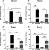
Levels of NO measured in blood (A), heart (B), liver (C), and lung (D) from MLCK210+/+ and MLCK210−/− mice injected with vehicle or LPS. Bars represent mean ± SEM (n = 3 or 6 per group): *P < 0.05, **P < 0.01, and ***P < 0.001 by one-way analysis of variance followed by Newman-Keuls post hoc test.

References

    1. Hotchkiss RS, Karl IE. The pathophysiology and treatment of sepsis. N Engl J Med. 2003;348:138–150. - PubMed
    1. Parrillo JE. Pathogenetic mechanisms of septic shock. N Engl J Med. 1993;328:1471–1478. - PubMed
    1. Court O, Kumar A, Parrillo JE, Kumar A. Clinical review: myocardial depression in sepsis and septic shock. Crit Care. 2002;6:500–508. - PMC - PubMed
    1. Dudek SM, Garcia JG. Cytoskeletal regulation of pulmonary vascular permeability. J Appl Physiol. 2001;91:1487–1500. - PubMed
    1. Yuan SY, Wu MH, Ustinova EE, Guo M, Tinsley JH, De Lanerolle P, Xu W. Myosin light chain phosphorylation in neutrophil-stimulated coronary microvascular leakage. Circ Res. 2002;90:1214–1221. - PubMed

Publication types

MeSH terms