. 2007 Feb;170(2):557-66.
doi: 10.2353/ajpath.2007.060893.

Exacerbated pathology of viral encephalitis in mice with central nervous system-specific autoantibodies

Affiliations

Exacerbated pathology of viral encephalitis in mice with central nervous system-specific autoantibodies

Renaud Burrer et al. Am J Pathol. 2007 Feb.

Abstract

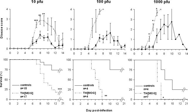
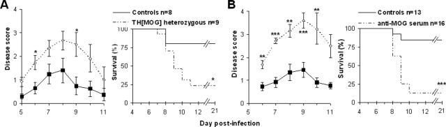
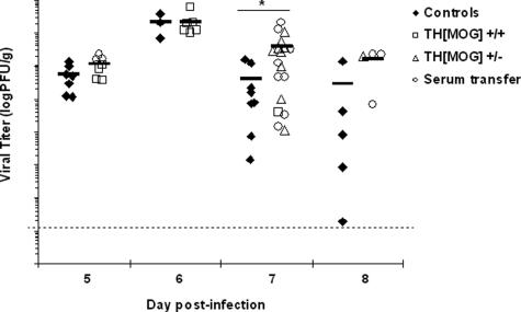
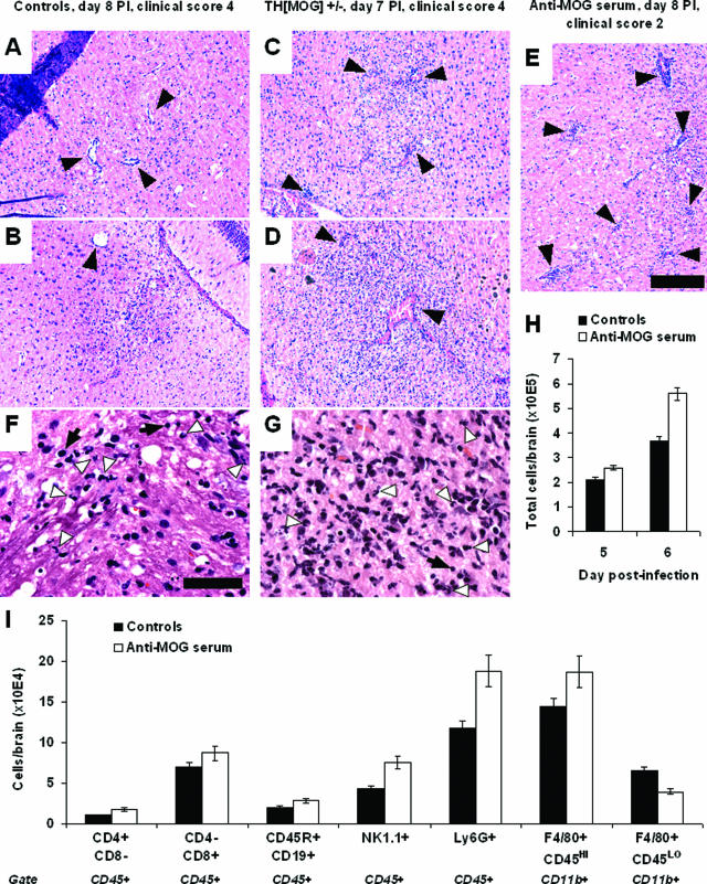
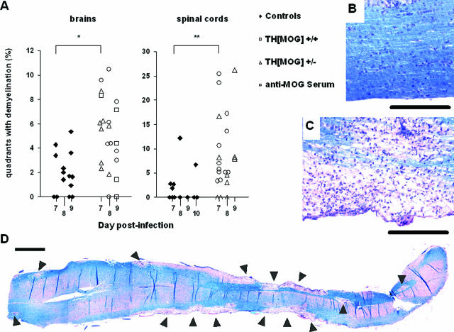
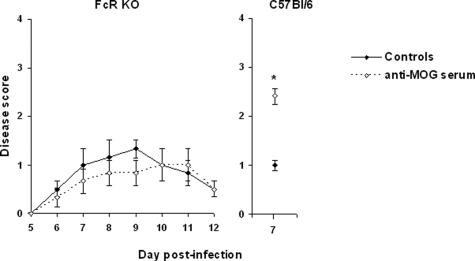
We examine here the outcome of viral encephalomyelitis [mouse hepatitis virus (MHV) A59, Theiler's encephalomyelitis virus, and Coxsackievirus B3] in mice with autoantibodies to a central nervous system (CNS)-specific antigen, myelin oligodendrocyte glycoprotein, that usually develop no clinical disease. Morbidity and mortality of the acute viral CNS disease was augmented by the presence of the autoantibodies in all three viral infections. Transfer of serum containing the autoantibodies at the time of infection with MHV was sufficient to reproduce the exacerbated disease. The presence of the autoantibodies was found to result in increased infiltration of mononuclear cells into the brain. Early demyelination was severely augmented in brains and spinal cords of MHV-infected mice with CNS-specific autoantibodies. The antibody-mediated exacerbation was shown to be independent of the complement system but to require expression of Fc receptors, because it was observed in C'-3-deficient but not in Fc receptor-deficient mice. Our study illustrates the possibility that infections can lead to much more profound immunopathology in the presence of an otherwise latent autoimmune condition.


Figures

Figure 1
Figure 1
The outcome of MHV A59 acute infection of the CNS is exacerbated in THMOG knock-in mice. Mice were observed and given a numerical score (top panels) for behavioral signs of encephalitis (0, no detectable sign of disease; 1, ruffled fur; 2, slightly hunched back and ruffled fur; 3, very hunched back and lethargy; and 4, death) daily during the acute infection phase in mice infected with increasing doses of virus (left to right). Data represent the average and standard errors for each group. The bottom panels show the survival of the animals in the same experiments. *P < 0.05; **P < 0.01; ***P < 0.001.
Figure 2
Figure 2
Autoantibodies account for the exacerbated viral encephalitis. A: Clinical disease and survival after intracranial (i.c.) inoculation of 10 PFU of MHV A59 in C57Bl/6 controls (black squares, plain lines) and THMOG heterozygous mice (open diamonds, dashed lines). B: Comparison of the viral encephalitis after injection of 10 PFU of MHV A59 in C57Bl/6 that received serum of THMOG mice intraperitoneally at the time of infection (open diamonds, dashed lines) or mock-treated controls (black squares, plain lines). *P < 0.05; **P < 0.01; ***P < 0.001.
Figure 3
Figure 3
Infectious virus levels in the brain. Brains obtained from control mice (black diamonds) or animals with autoantibodies (white squares, homozygous THMOG; white triangles, heterozygous THMOG; white circles, wild-type recipients of THMOG serum) infected with 10 PFU of MHV A59 were obtained at various time points after inoculation. Viral titers from homogenized half-brains were determined by plaque assay. Data are pooled from six independent experiments. Each point represents an individual animal, and black lines represent the average for each group. *P < 0.05.
Figure 4
Figure 4
Infiltration is augmented in the presence of anti-MOG antibodies. A–E: Representative H&E-stained sections from brains of MHV A59-infected mice. Moderate inflammation, with some perivascular cuffing (black arrows), was detected in the brains of control mice (A and B, both obtained from different moribund animals 8 days after the infection). The degree of infiltration was augmented in all mice with autoantibodies (E, recipient of anti-MOG serum at day 8, clinical score 2) or earlier in the course of infection (C and D, obtained from moribund mice at day 7 after infection). Black bar = 0.2 mm. F and G: Close-up pictures from sections A and D, respectively, showing mixed inflammatory cell infiltrates. White arrowheads indicate some individual polymorphonuclear cells, which also can be found in large cluster in G. Some cells with lymphocyte morphology (black arrows) can also be seen in infiltrates in both groups of mice. Black bar = 0.05 mm. H: Total numbers of mononuclear cells purified from individual brains collected from control (black bars) or recipient of anti-MOG serum (open bars) mice 5 or 6 days after infection with MHV A59. Average and SE, n = 6 (day 50) or 13/14 (day 6) per group. I: Brains from control mice (average clinical score, 0.56 ± 0.12, n = 8) and recipients of anti-MOG serum (1.78 ± 0.18, n = 7) were collected 6 days after MHV A59 infection. Mononuclear cells were purified from each individual brain, counted, pooled by group, and stained for flow cytometry analysis. The graph shows the average number of T lymphocytes (CD4+CD8-cd45+ and CD4-CD8+cd45+), B lymphocytes (B220+CD19+CD45+), NK cells (NK1.1+CD45+), neutrophils (Ly6G+CD45+), blood-borne macrophages (CD11b+F4/80+CD45HIGH), and resident microglia (CD11b+F4/80+CD45LOW) per animal. Similar results were obtained in additional independent experiments performed at days 5, 6, and 7 after infection.
Figure 5
Figure 5
Demyelinating lesions in the brains and spinal cords of MHV-A59-infected mice. Luxol Fast Blue-stained tissue sections from control and autoimmune mice were examined for demyelination. A: Quantification of demyelinating lesions in control (black diamonds) and autoimmune (empty symbols) mice. A square grid was applied to sagittal brain sections (left) and coronal spinal cord sections (right), and results are expressed as the percentage of the total quadrants displaying lesions in each individual animal. Differences were statistically significant at day 7 for both organs. *P < 0.05. **P < 0.01. B and C: Representative pictures of spinal cords (longitudinal sections) from a control animal (B) and a C57Bl/6 recipient of anti-MOG serum (C) both obtained at day 8 after infection with 10 PFU of MHV A59. Note the massive infiltration in the lesion in C. Black bars = 0.2 mm. D: The reconstituted picture shows demyelinating lesions (arrowheads) spreading over the entire length of a spinal cord longitudinal section, obtained from the same animal as in C. It is representative of mice with autoantibodies at 7 to 9 days after infection, whereas all of the control mice in this study had little or no lesions in the spinal cord. Black bar = 1 mm.
Figure 6
Figure 6
Anti-MOG antibodies do not exacerbate encephalitis in Fc receptor-deficient mice. Although all mice showed clinical signs of CNS infection, the intensity of MHV-induced disease was not affected by the transfer of anti-MOG autoantibodies in mice deficient in Fc receptors, and all of the mice survived the experiment in both groups (left, n = 3/group). MHV A59-infected wild-type mice transferred with the same serum pool displayed the normal exacerbation at the time they were sacrificed for a flow cytometry experiment (right, n = 6/group). *P < 0.05. The experiment was repeated as indicated in the main text section, confirming the absence of antibody-mediated pathology in the Fc receptor-deficient mice.

Comment in

References

    1. von Herrath MG, Fujinami RS, Whitton JL. Microorganisms and autoimmunity: making the barren field fertile? Nat Rev Microbiol. 2003;1:151–157. - PubMed
    1. Christen U, Edelmann KH, McGavern DB, Wolfe T, Coon B, Teague MK, Miller SD, Oldstone MB, von Herrath MG. A viral epitope that mimics a self antigen can accelerate but not initiate autoimmune diabetes. J Clin Invest. 2004;114:1290–1298. - PMC - PubMed
    1. Panoutsakopoulou V, Sanchirico ME, Huster KM, Jansson M, Granucci F, Shim DJ, Wucherpfennig KW, Cantor H. Analysis of the relationship between viral infection and autoimmune disease. Immunity. 2001;15:137–147. - PubMed
    1. McCoy L, Tsunoda I, Fujinami RS. Multiple sclerosis and virus induced immune responses: autoimmunity can be primed by molecular mimicry and augmented by bystander activation. Autoimmunity. 2006;39:9–19. - PubMed
    1. Buljevac D, Flach HZ, Hop WC, Hijdra D, Laman JD, Savelkoul HF, van Der Meche FG, van Doorn PA, Hintzen RQ. Prospective study on the relationship between infections and multiple sclerosis exacerbations. Brain. 2002;125:952–960. - PubMed

Publication types

MeSH terms