. 2007 Feb;170(2):774-86.
doi: 10.2353/ajpath.2007.060761.

Tumor-induced sentinel lymph node lymphangiogenesis and increased lymph flow precede melanoma metastasis

Affiliations

Tumor-induced sentinel lymph node lymphangiogenesis and increased lymph flow precede melanoma metastasis

Maria I Harrell et al. Am J Pathol. 2007 Feb.

Abstract

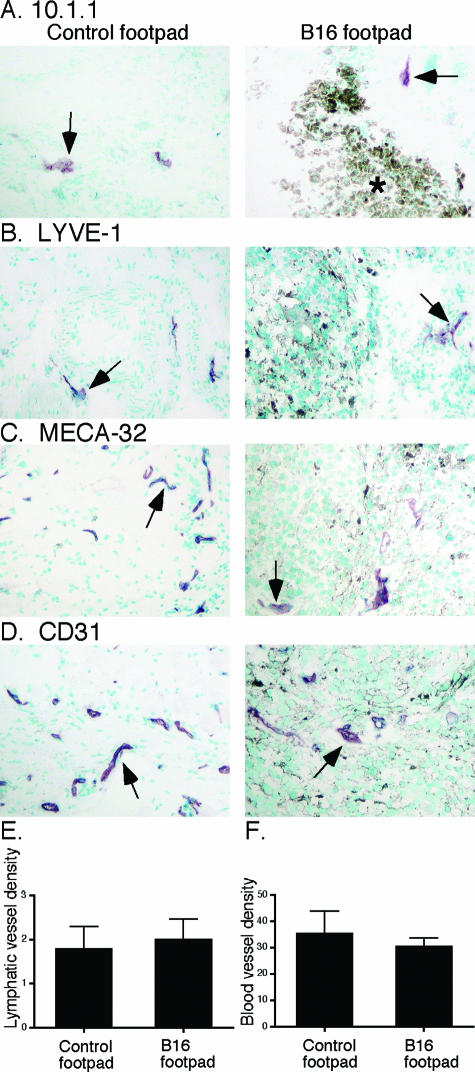
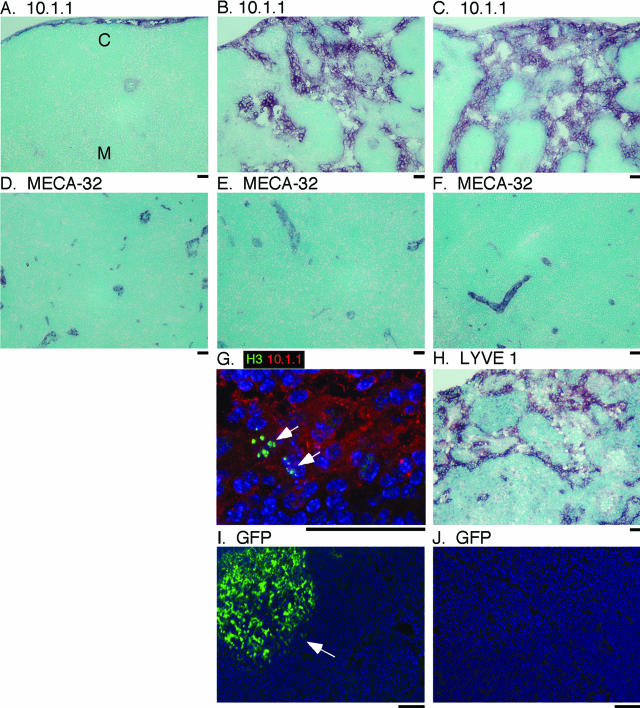
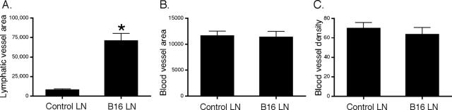
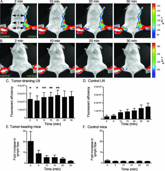
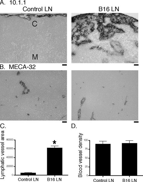
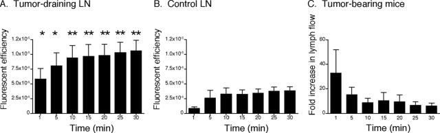
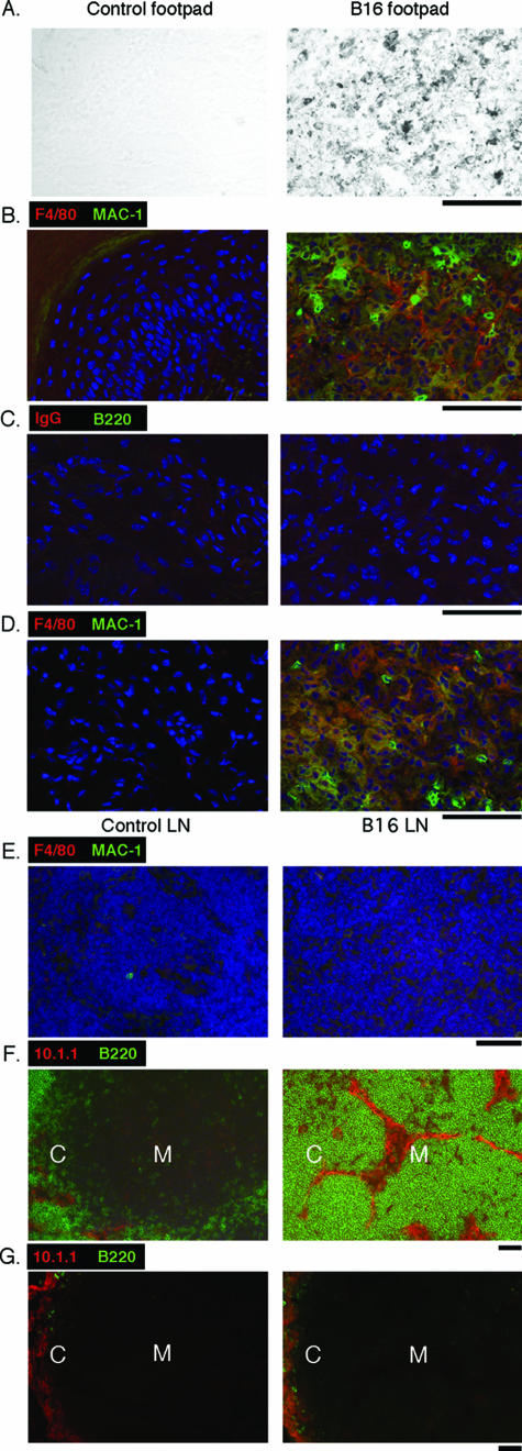
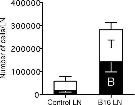
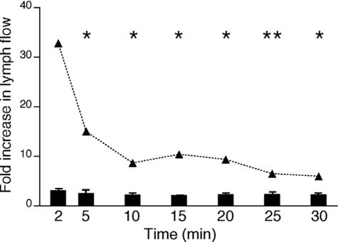
Lymphangiogenesis is associated with human and murine cancer metastasis, suggesting that lymphatic vessels are important for tumor dissemination. Lymphatic vessel alterations were examined using B16-F10 melanoma cells implanted in syngeneic C57Bl/6 mice, which form tumors metastasizing to draining lymph nodes and subsequently to the lungs. Footpad tumors showed no lymphatic or blood vessel growth; however, the tumor-draining popliteal lymph node featured greatly increased lymphatic sinuses. Lymph node lymphangiogenesis began before melanoma cells reached draining lymph nodes, indicating that primary tumors induce these alterations at a distance. Lymph flow imaging revealed that nanoparticle transit was greatly increased through tumor-draining relative to nondraining lymph nodes. Lymph node lymphatic sinuses and lymph flow were increased in mice implanted with unmarked or with foreign antigen-expressing melanomas, indicating that these effects are not due to foreign antigen expression. However, tumor-derived immune signaling could promote lymph node alterations, as macrophages infiltrated footpad tumors, whereas lymphocytes accumulated in tumor-draining lymph nodes. B lymphocytes are required for lymphangiogenesis and increased lymph flow through tumor-draining lymph nodes, as these alterations were not observed in mice deficient for B cells. Lymph node lymphangiogenesis and increased lymph flow through tumor-draining lymph nodes may actively promote metastasis via the lymphatics.


Figures

Figure 1
Figure 1
Normal representation of lymphatics and blood vessels in footpad melanomas in albino mice. A: Control footpad or B16 footpad tumor immunostained with 10.1.1 lymphatic endothelial antibody both show sparse purple-stained lymphatic vessels (arrows) with methyl green counterstaining. The black melanin pigment identifies the B16 tumor (right panel, asterisk). B: LYVE-1 immunostaining also identifies sparse lymphatic vessels (arrows) in control (left panel) or B16 footpads (right panel). C: Blood vessels immunostained with MECA-32 antibody (arrows) are similar in control footpads (left panel) and in footpad tumors (right panel). D: CD31 immunostaining also shows similar blood vessels in control and B16 footpads. All panels are shown at ×100 magnification. E: Image quantification demonstrates similar lymphatic vessel density in seven control and 10 B16 tumor-bearing footpads. F: Image quantification confirms similar blood vessel density in seven control and eight tumor-bearing footpads. Standard errors are shown.
Figure 2
Figure 2
Melanoma-draining LNs undergo lymphangiogenesis in albino mice. 10.1.1 immunostaining of wild-type LN shows few cortical (C) or medullary (M) lymphatic sinuses (A), whereas a tumor-draining popliteal LN containing a melanoma metastasis (B) or a LN that does not yet contain melanoma cells (C) shows extensive lymphangiogenesis throughout the cortex and medulla. Control (D), metastatic (E), or pre-metastatic (F) LNs show similar blood vessel density by MECA-32 immunostaining. Immunofluorescent staining for mitotic phosphohistone H3 detected with FITC-labeled secondary antibodies, and 10.1.1 lymphatic endothelial immunostaining with Alexa 594-labeled secondary antibodies shows mitotic lymphatic endothelium in tumor-draining LN (G, arrows). Nuclear DAPI staining is also shown. Immunohistochemical staining with LYVE-1 antibody confirms increased lymphatic sinuses in pre-metastatic tumor-draining LNs (H). Melanoma cells are detected in draining LNs by their GFP expression (I, arrow). Pre-metastatic LN does not contain GFP-positive cells (J). Scale bars = 25 μm.
Figure 3
Figure 3
Lymphangiogenesis in the tumor-draining LN is not associated with angiogenesis. Measurement of 10.1.1-positive lymphatic vessel area in seven control and seven tumor-draining leg popliteal LNs shows a ninefold increase in lymphatic vessels (A). This increase is statistically significant by t-test (N = 34, *P < 0.0001). In contrast, MECA 32-positive blood vessel area (B) and density (C) are similar in control and tumor-draining leg popliteal LNs (N = 36). Standard errors are shown.
Figure 4
Figure 4
Lymph flow increases in the tumor-draining LNs of albino mice. A: Quantum dots were injected into both dorsal rear toes of albino C57Bl/6 mice bearing tumors in the left rear footpad, and mice were imaged from 2 to 30 minutes after injection. Shown is a representative set of supine images. The popliteal LN in the tumor-draining leg (P, arrow) is positive for quantum dots within 2 minutes after injection, and the increased signal persists for 30 minutes relative to the control leg. The inguinal (I, arrow) and axillary (A, arrow) LNs draining the tumor are also positive. B: Control albino C57Bl/6 mouse injected with quantum dots shows no visible signal in either popliteal LN until 30 minutes after injection. C: Measurement of quantum dot or Cy5.5 nanoparticle fluorescent efficiency in a region of interest drawn over the tumor-draining popliteal LN from eight tumor-bearing mice. Lymph flow is significantly increased in the tumor-draining LN relative to the control nondraining LN by paired t-test. *P < 0.04, **P < 0.02. D: Fluorescent efficiency slowly increases in popliteal LN draining the control leg of tumor-bearing mice. E: Pairwise comparison of fluorescent efficiency in the tumor-draining and control LNs from individual mice demonstrates increased lymph flow in tumor-draining LNs expressed as a fold increase in lymph flow over the control LNs. F: Control mice not bearing tumors showed no difference in lymph flow through each popliteal LN. Standard errors are shown.
Figure 5
Figure 5
Lymphangiogenesis in LNs draining tumors that do not express foreign antigen. A: Popliteal LNs from the control leg of black (melanin-positive) mice implanted with unmarked (GFP-negative) tumors show few cortical (C) or medullary (M) lymphatic sinuses with 10.1.1 immunostaining (left panel), whereas B16 tumor-draining popliteal LN shows extensive growth of lymphatic sinuses throughout the cortex and medulla (right panel). B: MECA-32-positive blood vessels are equally abundant in control (left panel) or tumor-draining (right panel) LNs. Scale bars = 25 μm. C: Quantification of lymphatic vessel area demonstrates a 12-fold increase in lymphatic vessels in six tumor-draining LNs versus six control LNs, which is statistically significant by t-test (*P < 0.0001, N = 24). D: Blood vessel density is equal in tumor-draining LNs compared with control LNs (N = 16). Standard errors are shown.
Figure 6
Figure 6
Lymph flow increases in the tumor-draining LN of black mice. Black C57/Bl6 mice implanted with unmarked B16 tumors were imaged after quantum dot injection into both rear dorsal toes, and quantum dot fluorescent efficiency was measured in regions of interest over the popliteal LN in the tumor-draining and control legs from 10 tumor-bearing mice. Quantum dot accumulation is increased in the B16 tumor-draining popliteal LN (A) relative to the nondraining control leg LN (B) from 2 through 30 minutes of imaging, and this difference is statistically significant (*P < 0.04, **P < 0.03). Pairwise comparison of lymph flow through the tumor-draining versus control LNs from individual mice (C) demonstrates increased lymph flow through the tumor-draining LNs. Standard errors are shown.
Figure 7
Figure 7
Immune cell accumulation in the primary tumor and tumor-draining LN. A: Light microscopy of control (left panel) or B16-implanted (right panel) footpads identifies black-pigmented melanoma cells restricted to the B16 footpad. B: Immunostaining of the same footpad regions with Alexa 568-labeled F4/80 and FITC-labeled Mac-1 antibodies shows extensive accumulation of F4/80 and/or Mac-1-positive macrophages and other leukocytes throughout the footpad tumor (right panel) in wild-type mice, whereas these cells were absent from the control footpad (left panel). Nuclei are stained with DAPI. C: FITC-labeled B220 immunostaining shows that there are no B lymphocytes in control or B16 footpads of wild-type mice. Control Alexa 568-labeled anti-rat IgG antibodies show no immunostaining of footpads. D: Alexa 568-labeled F4/80 and FITC-labeled Mac-1-positive cells are abundant in footpad tumors (right panel) but not in control footpads (left panel) from μMT B cell-deficient mice. E: Control (left panel) or tumor-draining (right panel) LNs from wild-type mice show few Alexa 568-labeled F4/80 or FITC-labeled Mac-1-positive cells. F: FITC-labeled B220-positive lymphocytes and Alexa 568-labeled 10.1.1-positive lymphatic sinuses are restricted to the cortex (C) of control LN (left panel), whereas B16 tumor-draining LN features B220-positive B lymphocyte accumulation throughout the cortex and medulla (M) alongside enlarged 10.1.1-positive lymphatic sinuses (right panel). G: LN from μMT mice do not contain FITC-labeled B220-positive B lymphocytes, and Alexa 568-labeled 10.1.1-positive lymphatic sinuses are restricted to the cortex of both control (left panel) or tumor-draining (right panel) LNs. Scale bars = 50 μm.
Figure 8
Figure 8
Lymphocytes accumulate in tumor-draining LNs. B220-positive B lymphocytes (black bar) and CD3-positive T lymphocytes (white bar) from control or B16 LNs were measured by flow cytometry. B cell content was significantly increased in tumor-draining LNs, and this change was statistically significant by t-test (N = 3, P < 0.05).
Figure 9
Figure 9
Normal lymph flow through tumor-draining LN in μMT B lymphocyte-deficient mice. Quantum dots were injected into both feet of tumor-bearing μMT mice, and regions of interest were quantitated over 30 minutes of Xenogen imaging in four mice. Lymph flow through the tumor-draining LN was not significantly increased relative to the control leg LN (black bars), in contrast to the large increase in lymph flow through the tumor-draining LNs of wild-type mice (data from Figure 6C shown as a dashed line). This difference in lymph flow through the tumor-draining LN of μMT and wild-type mice is statistically significant by t-test (*P < 0.04, **P < 0.02).

References

    1. Cao Y. Emerging mechanisms of tumour lymphangiogenesis and lymphatic metastasis. Nat Rev Cancer. 2005;5:735–743. - PubMed
    1. He Y, Karpanen T, Alitalo K. Role of lymphangiogenic factors in tumor metastasis. Biochim Biophys Acta. 2004;1654:3–12. - PubMed
    1. Stacker S, Baldwin M, Achen M. The role of tumor lymphangiogenesis in metastatic spread. FASEB J. 2002;16:922–934. - PubMed
    1. Stacker SA, Caesar C, Baldwin ME, Thornton GE, Williams RA, Prevo R, Jackson DG, Nishikawa S, Kubo H, Achen MG. VEGF-D promotes the metastatic spread of tumor cells via the lymphatics. Nat Med. 2001;7:186–191. - PubMed
    1. Skobe M, Hawighorst T, Jackson DG, Prevo R, Janes L, Velasco P, Riccardi L, Alitalo K, Claffey K, Detmar M. Induction of tumor lymphangiogenesis by VEGF-C promotes breast cancer metastasis. Nat Med. 2001;7:192–198. - PubMed

Publication types