. 2007 Jan 26;315(5811):518-21.
doi: 10.1126/science.1134910.

Asymmetric inheritance of mother versus daughter centrosome in stem cell division

Affiliations

Asymmetric inheritance of mother versus daughter centrosome in stem cell division

Yukiko M Yamashita et al. Science. .

Abstract

Adult stem cells often divide asymmetrically to produce one self-renewed stem cell and one differentiating cell, thus maintaining both populations. The asymmetric outcome of stem cell divisions can be specified by an oriented spindle and local self-renewal signals from the stem cell niche. Here we show that developmentally programmed asymmetric behavior and inheritance of mother and daughter centrosomes underlies the stereotyped spindle orientation and asymmetric outcome of stem cell divisions in the Drosophila male germ line. The mother centrosome remains anchored near the niche while the daughter centrosome migrates to the opposite side of the cell before spindle formation.


Figures

Fig. 1
Fig. 1
GFP-labeled daughter centrosomes migrate away from the niche. (A) Stereotyped positioning of centrosomes in male GSC during inter-phase sets up the orientation of the mitotic spindle [adapted from (6)]. Red, centrosome; blue, hub; green, tubulin. (B) Pulse-chase strategy to label the daughter centrosome. Each centrosome (circle) contains two centrioles (ovals numbered in order of generation). Transient expression of GFP-PACT labels newly assembled centrioles (3). In the first cell cycle after the GFP-PACT pulse, both the mother (1+3) and daughter (2+3) centrosomes are GFP-positive. In the second cell cycle, the mother centrosomes (1+4 or 2+4) are GFP-negative, whereas the daughter centrosomes (3+4) are GFP-positive. (C and D) Testis tips with labeled GSCs (dotted outline) in the G2 phase of the first cell cycle after the GFP-PACT pulse [(C), 12 hours after heat shock] or the second cell cycle [(D), 24 hours after heat shock]. Green, GFP-PACT; red, γ-tubulin (centrosomes are shown with arrowheads) and Fas III (hub, H); blue, Vasa (germ cells). Scale bar, 10 μm. (E) Frequency of label outcomes over time after heat shock. Blue, GSCs in the first cell cycle (two labeled, oriented centrosomes); red, GSCs in the second cell cycle (one labeled and one unlabeled centrosome) with the labeled (daughter) centrosome distal from the hub. Rarely observed outcomes were pink, the first cell cycle, with neither centrosome next to the hub; black, the second cell cycle, with the mother centrosome distal to the hub; green, the second cell cycle, with neither centrosome next to the hub. Only those GSCs that had two centrosomes, with one or both centrosomes labeled with GFP-PACT, were counted.
Fig. 2
Fig. 2
Male GSCs in the niche maintain centrioles assembled many cell generations earlier. (A) Strategy to label the mother centrosome: GFP-PACT expressed during early embryogenesis using the NGT40 driver marks the centrioles (1 and 2) retained in the mother centrosomes after the depletion of cytoplasmic GFP-PACT. (B) Testis tip from a newly eclosed male with GSCs containing GFP-labeled mother centrosomes proximal to the hub-GSC interface. Red, γ-tubulin (centrosomes are shown with arrowheads) and Fas III (hub, H); green, GFP-PACT; blue, Vasa (germ cells). Scale bar, 10 μm. (C) Frequency of label outcomes over time from NGT40/UAS-GFP-PACT flies. UAS, upstream activating sequence. Blue columns, GSCs in the second or later cell cycle after GFP-PACT depletion (only one centrosome was labeled with GFP). 1 proximal, labeled centrosome located proximal to the hub-GSC interface; 1 distal, labeled centrosome located distal from the hub-GSC interface; 1 misori, neither of the two centrosomes located near the hub-GSC interface. Red columns, GSCs in the first cell cycle (both centrosomes were labeled by GFP). 2 ori, one centrosome close to the hub-GSC interface; 2 misori, neither centrosome close to the hub-GSC interface. Green column, GSCs that still had cytoplasmic GFP-PACT. Only 50 to 60% of the total male GSCs were labeled by NGT40/GFP-PACT. The remaining 40 to 50% could contain mother centrioles assembled in early embryogenesis before GFP-PACT expression. Only those GSCs that had two centrosomes, with one or both centrosomes labeled with GFP-PACT, were counted. N, number of GSCs scored per time point.
Fig. 3
Fig. 3
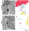
Centrosomes next to the hub harbor robust microtubule arrays. (A) Electron micrograph and (A′) summary diagram of a proximal centrosome in a GSC. Arrowheads in (A′) show a microtubule that runs from the centrosome to the adherens junction. (B) Electron micrograph and (B′) summary diagram of a distal centrosome in a GSC. Red, hub; blue, microtubules; green, adherens junctions; yellow, nucleus; pink, centriole; gray in (B′), plasma membrane. Scale bar, 1 μm.
Fig. 4
Fig. 4
cnn is required for nonrandom segregation of mother and daughter centrosomes. (A) Summary of the centrosome-positioning pattern in cnnHK21 homozygous mutant and control cnnHK21/+ GSCs. Daughter centrosomes were labeled by a pulse of GFP-PACT as in Fig. 1B. Only counts of cells in the second cell cycle are shown. (B and C) Testis tips from cnn males with GSCs in the second cell cycle with misoriented centrosomes (B) or with the mother rather than the daughter centrosome segregated to the opposite side of the GSC (C). Red, γ-tubulin [centrosomes are shown with arrowheads (M, mother; D, daughter)] and Fas III (hub, H); green, GFP-PACT; blue, Vasa (germ cells). Scale bar, 10 μm.

Comment in

Similar articles

Cited by

References

    1. Morrison SJ, Kimble J. Nature. 2006;441:1068. - PubMed
    1. Spradling A, Drummond-Barbosa D, Kai T. Nature. 2001;414:98. - PubMed
    1. Watt FM, Hogan BLM. Science. 2000;287:1427. - PubMed
    1. Kiger AA, Jones DL, Schulz C, Rogers MB, Fuller MT. Science. 2001;294:2542. - PubMed
    1. Tulina N, Matunis E. Science. 2001;294:2546. - PubMed

Publication types

MeSH terms

Substances