Comparative Study
. 2007 Apr 15;175(8):805-15.
doi: 10.1164/rccm.200608-1162OC. Epub 2007 Jan 25.

Nuclear factor-kappaB activation in neonatal mouse lung protects against lipopolysaccharide-induced inflammation

Affiliations
Comparative Study

Nuclear factor-kappaB activation in neonatal mouse lung protects against lipopolysaccharide-induced inflammation

Cristina M Alvira et al. Am J Respir Crit Care Med. .

Abstract

Rationale: Injurious agents often cause less severe injury in neonates as compared with adults.

Objective: We hypothesized that maturational differences in lung inflammation induced by lipopolysaccharide (LPS) may be related to the nature of the nuclear factor (NF)-kappaB complex activated, and the profile of target genes expressed.

Methods: Neonatal and adult mice were injected with intraperitoneal LPS. Lung inflammation was assessed by histology, and apoptosis was determined by TUNEL (terminal deoxynucleotidyl transferase UTP nick-end labeling). The expression of candidate inflammatory and apoptotic mediators was evaluated by quantitative real-time polymerase chain reaction and Western immunoblot.

Results: Neonates demonstrated reduced inflammation and apoptosis, 24 hours after LPS exposure, as compared with adults. This difference was associated with persistent activation of NF-kappaB p65p50 heterodimers in the neonates in contrast to early, transient activation of p65p50 followed by sustained activation of p50p50 in the adults. Adults had increased expression of a panel of inflammatory and proapoptotic genes, and repression of antiapoptotic targets, whereas no significant changes in these mediators were observed in the neonates. Inhibition of NF-kappaB activity in the neonates decreased apoptosis, but heightened inflammation, with increased expression of the same inflammatory genes elevated in the adults. In contrast, inhibition of NF-kappaB in the adults resulted in partial suppression of the inflammatory response.

Conclusions: NF-kappaB activation in the neonatal lung is antiinflammatory, protecting against LPS-mediated lung inflammation by repressing similar inflammatory genes induced in the adult.


Figures

<b>Figure 1.</b>
Figure 1.
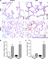
LPS induces increased lung inflammation in adult versus neonatal mice. (A) Representative formalin-fixed lung sections stained by hematoxylin and eosin demonstrate increased inflammatory cell infiltration, alveolar thickening, and edema in the adult lung in contrast to little gross histologic change observed in the neonatal lung (Neo) 24 hours after LPS exposure. Immunostaining of representative sections demonstrates increased (B) Mac-3– and (C) Ly 7/4–positive cells in LPS-treated adult lungs as compared with neonatal lungs at 24 hours. (D) Quantification of Mac-3– and Ly 7/4–positive cells per field in control (Con) and LPS-treated neonatal and adult mice. Bars represent mean ± SEM for control (n = 3) and LPS-treated adult and neonatal mice (n = 4 for both). **p < 0.01 and ***p < 0.001 versus control adults. Scale bar represents 50 μm.
<b>Figure 2.</b>
Figure 2.
LPS induces increased apoptosis in adult versus neonatal mice. (A) Representative TUNEL staining of formalin-fixed lung sections demonstrates increased positive cells in adult mice in response to LPS exposure. (B) Quantification of TUNEL-positive cells from five representative lung sections in a blinded fashion demonstrates increased apoptotic cells in neonatal lungs under control conditions. Both LPS-treated adults and neonates had increased TUNEL-positive cells as compared with control animals. Bars represent mean ± SEM for control (n = 3) and LPS-treated adult and neonatal mice (n = 4 for both). *p < 0.05 versus control neonates and **p < 0.01 versus control adults. (C) A greater fold increase was seen in the number of apoptotic cells in the LPS-treated adults as compared with the LPS-treated neonates. *p = 0.01 versus LPS treated neonates. Scale bar represents 50 μm.
<b>Figure 3.</b>
Figure 3.
Systemic LPS activates distinct nuclear factor (NF)-κB complexes in adult and neonatal mouse lungs. (A) Nuclear extracts from lung homogenates were incubated with radiolabeled oligonucleotides containing the NF-κB consensus sequence. Activated NF-κB complexes were present in the nuclear extracts of both LPS-treated neonatal and adult mice, beginning at 1 hour (lanes 3 and 11), peaking at 2 hours (lanes 4, 5, 12, 13) and decreasing by 4 hours (lanes 6 and 14). However, the primary complex in the neonates (lanes 4 and 5) was noted to have a slower speed of migration as compared with the primary complex found in the adults (lanes 12 and 13). Specificity of the bands was confirmed by the disappearance of the bands with the addition of 100-fold excess of cold oligonucleotide (Cold oligo) (lanes 7 and 15), and the reappearance of the bands with the addition of 100-fold excess of cold, mutated oligonucleotide (Mutant oligo) (lanes 8 and 16). Supershift analysis of the neonatal (B) and adult (C) NF-κB complexes present at 2 hours was performed by the addition of antibodies against the Rel family proteins: p65, p50, Rel B, and cRel. There is an upward shift of the neonatal NF-κB complex with the addition of antibodies against p50, and decreased intensity of the band with the addition of antibodies against p65. Supershift analysis of the adult complex demonstrates a shift in the position or intensity of the band only by the addition of p50 antibodies. Images are representative examples of n ⩾ 3 individual experiments.
<b>Figure 4.</b>
Figure 4.
Increased nuclear translocation of p65 NF-κB in LPS-treated neonates compared with adults. Representative immunostaining for the p65 subunit in formalin-fixed lung sections demonstrates abundant cytoplasmic staining of p65 in both adult and neonatal mice under control conditions. Increased nuclear translocation of p65 was found in the neonatal lung 2 hours after LPS exposure as compared with minimal p65 nuclear translocation in the adult lung. Scale bar represents 50 μm. Images are representative examples from groups of adult and neonatal control (n = 3) and LPS-treated (n = 4) mice.
<b>Figure 5.</b>
Figure 5.
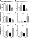
Decreased antiapoptotic and increased proapoptotic factors in LPS-treated adult mice. (A, B) Western immunoblot analysis demonstrated decreased optical density (OD) cellular inhibitor of apoptosis-1 (cIAP-1) (A) and cellular FLICE inhibitory protein (cFLIP) (B) levels in LPS-treated adults compared with LPS-treated neonates at 24 hours. Bars represent mean ± SEM for control and LPS-treated adult and neonatal mice (n = 3 for each group). **p < 0.01 and ***p < 0.001 versus control adults. (C) At 6 hours, higher basal expression of murine double minute-2 protein (Mdm2) in the neonates compared with adults under control conditions, and decreased expression of Mdm2 in the LPS-treated adults versus LPS-treated neonates. Bars represent mean ± SEM for control and LPS-treated adult and neonatal mice (n = 3 for each group). #p < 0.05 versus control neonates and **p < 0.01 versus LPS-treated neonates. (DF) LPS mediated up-regulation of the p53-dependent genes Noxa (D), DR5 (E), and Fas (F) in adult animals at 24 hours, in contrast to no significant elevation of these mediators in the neonates at this time point. Bars represent mean ± SEM for control and LPS-treated adult and neonatal mice (n = 3 for each group). *p < 0.05 versus control adults.
<b>Figure 6.</b>
Figure 6.
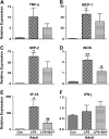
Increased expression of proinflammatory cytokines and chemokines in LPS-treated adult mice. Increased expression of tumor necrosis factor (TNF)-α (A), monocyte chemoattractant protein (MCP)-1 (B), macrophage inflammatory protein (MIP)-2 (C), inducible nitric oxide synthase (iNOS) (D), and interferon-γ–inducible protein-10 (IP-10) (E) observed in adult lungs, 24 hours after LPS exposure, in contrast to no significant increase in these mediators in LPS neonatal lungs. Neither adult nor neonatal lungs demonstrated statistically significant increases in the expression of IFN-γ (F) in response to LPS exposure. Bars represent mean ± SEM for control and LPS-treated adult and neonatal mice (n = 3 for each group). *p < 0.05 and ***p < 0.001 versus control adults.
<b>Figure 7.</b>
Figure 7.
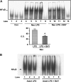
Administration of the selective NF-κB inhibitor BAY 11-7082 decreases NF-κB binding in vivo. Nuclear extracts obtained from neonates treated with LPS alone, or BAY 11-7082 before LPS, were used to evaluate the amount of NF-κB–DNA binding by electrophoretic mobility shift assay (EMSA). (A) Increased binding of NF-κB complexes in the LPS-treated neonates (lanes 4–7) 2 hours after exposure, as compared with controls (lanes 1–3). Administration of BAY 11-7082 1 hour before LPS exposure (lanes 9–12) decreased the NF-κB band intensity. Specificity of the bands was demonstrated by the disappearance of the band with the addition of 100-fold excess of cold oligonucleotide (lanes 8 and 13). Quantification of the bands shown in (A) by densitometry revealed a 42% reduction in the degree of NF-κB–DNA binding by the administration of BAY 11-7082 before LPS exposure. Bars represent mean ± SEM for control (n = 3), LPS-treated (n = 4), or BAY 11-7082 + LPS–treated neonates (n = 4), with *p = 0.008 versus LPS treated neonates by Student's t test. (B) EMSA using nuclear extracts obtained from adults treated with LPS alone (lanes 1–3), or with BAY 11-7082 before LPS (lanes 5–7), demonstrates that BAY 11-7082 does not decrease the DNA binding of the p50p50 complex.
<b>Figure 8.</b>
Figure 8.
Inhibition of NF-κB suppresses LPS-induced apoptosis in the neonatal lung. TUNEL staining of formalin-fixed lung sections 24 hours after LPS exposure demonstrates decreased TUNEL-positive cells in neonatal mice treated with the NF-κB inhibitor BAY 11-7082 as compared with neonates receiving LPS alone. Bars represent mean ± SEM for control (n = 3), LPS-treated (n = 4), or BAY 11-7082 + LPS–treated (n = 4) neonates. *p < 0.05 versus LPS treated neonates. Scale bar represents 50 μm.
<b>Figure 9.</b>
Figure 9.
Inhibition of NF-κB increases LPS-induced inflammation in the neonatal lung. (A) Hematoxylin and eosin staining of formalin-fixed lung sections demonstrates increased inflammatory cell infiltration and alveolar thickening in neonates treated with BAY 11-7082 as compared with neonates treated with LPS alone. (B, C) Quantification of (B) Mac-3– and (C) Ly 7/4–positive cells by immunostaining of representative sections demonstrates increased macrophages and neutrophils in the lungs of neonates treated with NF-κB inhibition as compared with neonates given LPS alone. Bars represent mean ± SEM for control (n = 3), LPS-treated (n = 4), or BAY 11-7082 + LPS–treated (n = 4) animals, with *p < 0.05 and **p < 0.01 versus LPS-treated neonates. Scale bar represents 50 μm.
<b>Figure 10.</b>
Figure 10.
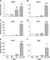
Inhibition of NF-κB increases inflammatory gene expression in LPS-treated neonates. Quantitative reverse transcriptase–polymerase chain reaction at 24 hours demonstrated increased expression of TNF-α (A), MCP-1 (B), MIP-2 (C), iNOS (D) interferon-γ–inducible protein-10 (IP-10) (E), and IFN-γ (F) in neonates treated with BAY 11-7082 before LPS as compared with LPS alone. The administration of BAY 11-7082 in the absence of LPS did not induce inflammatory gene expression. Bars represent mean ± SEM for control (n = 3), LPS-treated (n = 3), or BAY 11-7082 + LPS–treated (n = 3) animals. #p < 0.05 versus control neonates, and *p < 0.05, **p < 0.01, and ***p < 0.001 versus control, BAY alone, and LPS-treated neonates.
<b>Figure 11.</b>
Figure 11.
Effect of BAY 11-7082 on inflammatory gene expression in the LPS-treated adults. Quantitative reverse transcriptase–polymerase chain reaction at 24 hours in the adult animals treated with BAY before LPS as compared with LPS alone demonstrates no significant effect on the expression of TNF-α (A), MCP-1 (B), MIP-2 (C), and IFN-γ (F). However, BAY 11-7082 administration did reduce the expression of iNOS (D) and interferon-γ–inducible protein-10 (IP-10) (E) in the LPS-treated adults, providing further evidence of the proinflammatory role of NF-κB in the adult mice. Bars represent mean ± SEM for control (n = 3), LPS-treated (n = 3), or BAY 11-7082 + LPS–treated (n = 3) animals. *p < 0.05 and **p < 0.01 versus control adults, and #p < 0.05 and ##p < 0.01 versus LPS-treated adults.

References

    1. Piantadosi CA, Schwartz DA. The acute respiratory distress syndrome. Ann Intern Med 2004;141:460–470. - PubMed
    1. Estenssoro E, Dubin A, Laffaire E, Canales H, Saenz G, Moseinco M, Pozo M, Gomez A, Baredes N, Jannello G, et al. Incidence, clinical course, and outcome in 217 patients with acute respiratory distress syndrome. Crit Care Med 2002;30:2450–2456. - PubMed
    1. Rocco TR Jr, Reinert SE, Cioffi W, Harrington D, Buczko G, Simms HH. A 9-year, single-institution, retrospective review of death rate and prognostic factors in adult respiratory distress syndrome. Ann Surg 2001;233:414–422. - PMC - PubMed
    1. Matute-Bello G, Winn RK, Jonas M, Chi EY, Martin TR, Liles WC. Fas (CD95) induces alveolar epithelial cell apoptosis in vivo: implications for acute pulmonary inflammation. Am J Pathol 2001;158:153–161. - PMC - PubMed
    1. Matute-Bello G, Liles WC, Steinberg KP, Kiener PA, Mongovin S, Chi EY, Jonas M, Martin TR. Soluble Fas ligand induces epithelial cell apoptosis in humans with acute lung injury (ARDS). J Immunol 1999;163:2217–2225. - PubMed

Publication types