. 2007 Apr 15;175(8):829-39.
doi: 10.1164/rccm.200509-1527OC. Epub 2007 Jan 25.

Signal transduction pathways of tumor necrosis factor--mediated lung injury induced by ozone in mice

Affiliations

Signal transduction pathways of tumor necrosis factor--mediated lung injury induced by ozone in mice

Hye-Youn Cho et al. Am J Respir Crit Care Med. .

Abstract

Rationale: Increasing evidence suggests that tumor necrosis factor (TNF)-alpha plays a key role in pulmonary injury caused by environmental ozone (O(3)) in animal models and human subjects. We previously determined that mice genetically deficient in TNF response are protected from lung inflammation and epithelial injury after O(3) exposure.

Objectives: The present study was designed to determine the molecular mechanisms of TNF receptor (TNF-R)-mediated lung injury induced by O(3).

Methods: TNF-R knockout (Tnfr(-/-)) and wild-type (Tnfr(+/+)) mice were exposed to 0.3 ppm O(3) or air (for 6, 24, or 48 h), and lung RNA and proteins were prepared. Mice deficient in p50 nuclear factor (NF)-kappaB (Nfkb1(-/-)) or c-Jun-NH(2) terminal kinase 1 (Jnk1(-/-)) and wild-type controls (Nfkb1(+/+), Jnk1(+/+)) were exposed to O(3) (48 h), and the role of NF-kappaB and mitogen-activated protein kinase (MAPK) as downstream effectors of lung injury was analyzed by bronchoalveolar lavage analyses.

Results: O(3)-induced early activation of TNF-R adaptor complex formation was attenuated in Tnfr(-/-) mice compared with Tnfr(+/+) mice. O(3) significantly activated lung NF-kappaB in Tnfr(+/+) mice before the development of lung injury. Basal and O(3)-induced NF-kappaB activity was suppressed in Tnfr(-/-) mice. Compared with Tnfr(+/+) mice, MAPKs and activator protein (AP)-1 were lower in Tnfr(-/-) mice basally and after O(3). Furthermore, inflammatory cytokines, including macrophage inflammatory protein-2, were differentially expressed in Tnfr(-/-) and Tnfr(+/+) mice after O(3). O(3)-induced lung injury was significantly reduced in Nfkb1(-/-) and Jnk1(-/-) mice relative to respective control animals.

Conclusions: Results suggest that NF-kappaB and MAPK/AP-1 signaling pathways are essential in TNF-R-mediated pulmonary toxicity induced by O(3).


Figures
<b>Figure 1.</b>
Figure 1.
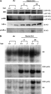
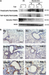
Intracellular tumor necrosis factor receptor (TNF-R) signal transducers were suppressed in Tnfr-deficient mice. (A) Differential activation of TNF-R proximal signaling complex in Tnfr+/+ and Tnfr−/− mice after exposure to air and 0.3 ppm O3 (6 or 24 h). Aliquots of total lung homogenates were immunoprecipitated (IP) using anti–TNF-R1–associated death domain protein (TRADD) or anti–TNF-R2 antibody followed by Western blot (WB) with anti–TNF-R–associated factor 2 (TRAF2) antibody to determine TRADD-bound TRAF2 or TNF-R2–bound TRAF2 levels, respectively. Soluble TRAF2 was determined by Western blot of nonparticulate fractions from lung homogenates. Representative images from multiple analyses (n = 3–4/group) are presented. Data are normalized to air-exposed Tnfr+/+ mice, and normalized group mean ± SEM and results from statistical analyses (two-way analysis of variance [ANOVA], p < 0.05) are shown in Table 1. (B) Differential levels of TRAF2 localized in terminal bronchioles of Tnfr+/+ and Tnfr−/− mice after 48 hours of exposure to air and 0.3 ppm O3. TRAF2-positive lung cells were immunohistologically stained with anti-TRAF2 antibody. High magnification shows TRAF2 localized on the membrane and in the cytoplasm. Bars indicate 100 μm. Representative light photomicrographs are presented.
<b>Figure 2.</b>
Figure 2.
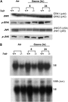
Nuclear factor (NF)-κB pathway was attenuated in Tnfr-deficient mice. (A) Differential activation of IKK and IκB in the lungs of Tnfr+/+ and Tnfr−/− mice after exposure to air and 0.3 ppm O3 (6 or 24 h) as assessed by Western blotting with phospho-specific antibodies. Representative images from multiple analyses (n = 3–4/group) are presented. Data are normalized to air-exposed Tnfr+/+ mice, and normalized group mean ± SEM and results from statistical analyses (two-way ANOVA, p < 0.05) are shown in Table 1. (B) Differential nuclear NF-κB–DNA binding activity in the lungs of Tnfr+/+ and Tnfr−/− mice after exposure to air and 0.3 ppm O3 (6 or 24 h). Aliquots of nuclear protein isolated from pieces of right lung (n = 3 mice/group) were incubated with an end-labeled oligonucleotide probe containing NF-κB consensus sequence. Total NF-κB–DNA binding was determined by gel shift analysis (top panel). To detect specific binding activity of each NF-κB subunit by gel supershift analyses, either anti-p65 (middle panel) or anti-p50 (bottom panel) subunit antibodies were added to the binding reactions. SB indicates shifted bands of total bindings (NF-κB motif–protein complex); SSB indicates super-shifted bands of specific bindings (NF-κB motif–protein antibody complex). Representative images from multiple analyses (n = 3/group) are presented. Data are normalized to air-exposed Tnfr+/+ mice, and normalized group mean ± SEM and results from statistical analyses (two-way ANOVA, p < 0.05) are shown in Table 1.
<b>Figure 3.</b>
Figure 3.
Mitogen-activated protein kinase/activator protein-1 (MAPK/AP-1) pathway was attenuated in Tnfr-deficient mice. (A) Differential phosphorylation of MAPK (p-JNK1, p-ERK) levels in the lungs of Tnfr+/+ and Tnfr−/− mice after exposure to air and 0.3 ppm O3 (6 or 24 h) as assessed by Western blotting. Representative images from multiple analyses (n = 3–4/group) are presented, and group mean ± SEM normalized to air-exposed Tnfr+/+ mice and statistical analyses are shown in Table 1. (B) Differential AP-1–DNA binding activity in the lungs of Tnfr+/+ and Tnfr−/− mice after air and 0.3 ppm O3 (6 or 24 h). Aliquots of nuclear protein isolated from pieces of right lung (n = 3 mice/group) were incubated with an end-labeled oligonucleotide probe containing AP-1 consensus sequence. Total AP-1–DNA binding was determined by gel shift analysis (top panel). To detect specific binding activity of AP-1 Jun proteins by gel supershift analyses, anti-Jun antibody was added to the binding reactions. SB indicates shifted bands of total bindings (AP-1 motif–protein complex); SSB indicates super-shifted bands of specific bindings (AP-1 motif–protein–antibody complex). Representative images from multiple analyses are presented. Data are normalized to air-exposed Tnfr+/+ mice, and normalized group mean ± SEM (n = 3/group) or mean and individual values (total AP-1 binding, n = 2/group) are shown with results from statistical analyses (two-way ANOVA, p < 0.05) in Table 1.
<b>Figure 4.</b>
Figure 4.
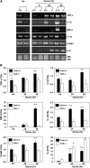
Tnfr deficiency reduced transcriptional induction of inflammatory mediators. Inflammatory gene expression was detected by reverse transcriptase–polymerase chain reaction using total lung RNA isolated from Tnfr+/+ and Tnfr−/− mice after exposure to air and 0.3 ppm O3 (n = 3/group). Representative cDNA band images for each gene are shown in (A). Quantitated intensities of digitized cDNA bands were normalized to the intensities of 18S bands, and relative intensity to air-exposed Tnfr+/+ mice of each gene is shown in (B). Data are presented as group means ± SEM. *Significantly higher than genotype-matched air controls (two-way ANOVA, p < 0.05); +significantly higher than exposure-matched Tnfr−/− mice (two-way ANOVA, p < 0.05). ICAM-1 = intercellular adhesion molecule-1; LT-β = lymphotoxin-β; MIP-2 = macrophage inflammatory protein-2; TNF-α = tumor necrosis factor-α.
<b>Figure 5.</b>
Figure 5.
Nuclear factor (NF)-κB was essential in O3-induced pulmonary pathogenesis. (A) Effect of targeted disruption of Nfkb1 was determined by bronchoalveolar lavage (BAL) phenotypes after 48 hours of exposure to air and 0.3 ppm O3. Data are presented as means ± SEM (n = 5 mice/group). *Significantly different from genotype-matched air control mice (two-way ANOVA, p < 0.05); +significantly different from O3-exposed Nfkb1+/+ mice (two-way ANOVA, p < 0.05). (B) Differential proliferation of pulmonary cells in Nfkb1+/+ and Nfkb1−/− mice after 48 hours of exposure to air or 0.3 ppm O3. S-phase cells undergoing proliferation were detected by proliferating cell nuclear antigen (PCNA) immunostaining. Representative light photomicrographs are shown. Bars indicate 100 μm. Representative Western blot image demonstrates NF-κB p50-dependent increase of nuclear PCNA in mouse lungs. Graph depicts mean ± SEM from duplicates normalized to air-exposed Nfkb+/+. *Significantly different from genotype-matched air control mice (two-way ANOVA, p < 0.05); +significantly different from exposure-matched Nfkb1+/+ mice (two-way ANOVA, p < 0.05).
<b>Figure 6.</b>
Figure 6.
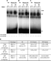
p50 Nuclear factor (NF)-κB deficiency abolished total NF-κB activity in the lung. Nuclear NF-κB–DNA binding activity in the lungs of Nfkb1+/+ and Nfkb1−/− mice after exposure to air and 0.3 ppm O3 (6 or 24 h). Aliquots of nuclear protein isolated from pieces of right lung (n = 3 mice/group) were incubated with an end-labeled oligonucleotide probe containing NF-κB consensus sequence. Total NF-κB–DNA binding was determined by gel shift analysis (A). To detect specific binding activity of each NF-κB subunit by gel supershift analyses, either anti-p50 (B) or anti-p65 (C) subunit antibody was added to the binding reactions. SB indicates shifted bands of total bindings (NF-κB motif–protein complex); SSB indicates super-shifted bands of specific bindings (NF-κB motif–protein–antibody complex); FP indicates free probes. The total (indicated as SB in A) and specific (indicated as SSB) p50 (B) or p65 (C) NF-κB–DNA binding activity was quantified using a Bio-Rad Gel Doc 2000 System, and mean ± SEM (n = 3/group) or mean and individual values (n = 2/group) normalized to air-exposed Nfkb1+/+ mice were presented (D). *Significantly different from genotype-matched air control mice (two-way ANOVA for total and p65 κB, one-way ANOVA for p50 κB, p < 0.05); +significantly different from exposure-matched Nfkb1+/+ mice (two-way ANOVA, p < 0.05).
<b>Figure 7.</b>
Figure 7.
c-Jun–NH2 terminal kinase (JNK) was essential in O3-induced pulmonary pathogenesis. Effect of targeted disruption of Jnk1 was determined by bronchoalveolar lavage (BAL) phenotypes after 48 hours of exposure to 0.3 ppm O3. Data are presented as means ± SEM (n = 3–5 mice/group). *Significantly different from genotype-matched air control mice (two-way ANOVA, p < 0.05); +significantly different from O3-exposed Jnk1+/+ mice (two-way ANOVA, p < 0.05).
<b>Figure 8.</b>
Figure 8.
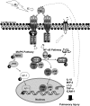
A hypothetical molecular mechanism underlying inhaled O3–induced pulmonary inflammation and injury. O3 may cause ligand binding to tumor necrosis factor receptor (TNF-R) on pulmonary cells to elicit trimerization of TNF-R and receptor complex formation by recruitment of accessory proteins, including TNF-R1–associated death domain protein (TRADD) and TNF-R–associated factor 2 (TRAF2). This event will trigger phosphorylation of downstream signal transducers, including mitogen-activated protein kinase (MAPK) kinase (MEK) and inhibitor of κB (IκB) kinase (IKK), which in turn would induce phosphorylation of MAPK, including c-Jun–NH2 terminal kinase (JNK) and phosphorylational degradation of IκB, respectively. Activator protein (AP)-1 proteins activated by phosphorylated MAPK and nuclear factor (NF)-κB subunits (e.g., p50, p65) liberated from IκB–NF-κB complex would be subsequently translocalized into nuclei for DNA binding to modulate inflammatory effector gene expression. These signaling pathways and possibly feedback regulation by TNF-α (dashed arrows) and/or by other cytokines and receptors (dotted arrow) may be essential to propagate airway inflammation and injury caused by O3, and exacerbate symptoms in subjects with preexisting respiratory disease (e.g., asthma).

Similar articles

Cited by

References

    1. Peel JL, Tolbert PE, Klein M, Metzger KB, Flanders WD, Todd K, Mulholland JA, Ryan PB, Frumkin H. Ambient air pollution and respiratory emergency department visits. Epidemiology 2005;16:164–174. - PubMed
    1. Tolbert PE, Mulholland JA, MacIntosh DL, Xu F, Daniels D, Devine OJ, Carlin BP, Klein M, Dorley J, Butler AJ, et al. Air quality and pediatric emergency room visits for asthma in Atlanta, Georgia, USA. Am J Epidemiol 2000;151:798–810. - PubMed
    1. Peden DB. Pollutants and asthma: role of air toxics. Environ Health Perspect 2002;110:565–568. - PMC - PubMed
    1. Vagaggini B, Taccola M, Cianchetti S, Carnevali S, Bartoli ML, Bacci E, Dente FL, Di Franco A, Giannini D, Paggiaro PL. Ozone exposure increases eosinophilic airway response induced by previous allergen challenge. Am J Respir Crit Care Med 2002;166:1073–1077. - PubMed
    1. Locksley RM, Killeen N, Lenardo MJ. The TNF and TNF receptor superfamilies: integrating mammalian biology. Cell 2001;104:487–501. - PubMed

Publication types

MeSH terms