. 2007 Jan;119(1):71-83.
doi: 10.1097/01.prs.0000244840.80661.e7.

Adipose tissue engineering from human adult stem cells: clinical implications in plastic and reconstructive surgery

Affiliations

Adipose tissue engineering from human adult stem cells: clinical implications in plastic and reconstructive surgery

Michael S Stosich et al. Plast Reconstr Surg. 2007 Jan.

Abstract

Background: Despite certain levels of clinical efficacy, current autografts and synthetic materials for soft-tissue reconstruction and/or augmentation suffer from donor-site morbidity, rupture, dislocation, and volume reduction. Human adult stem cells can self-replicate and differentiate into adipogenic cells in response to appropriate signaling cues. This study investigated the shape and dimension maintenance of engineered adipose tissue from adult human mesenchymal stem cells.

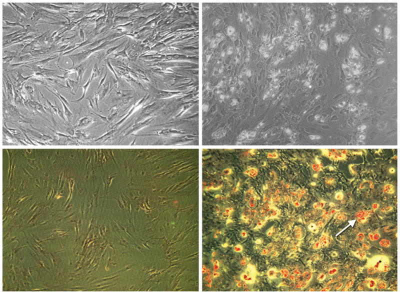
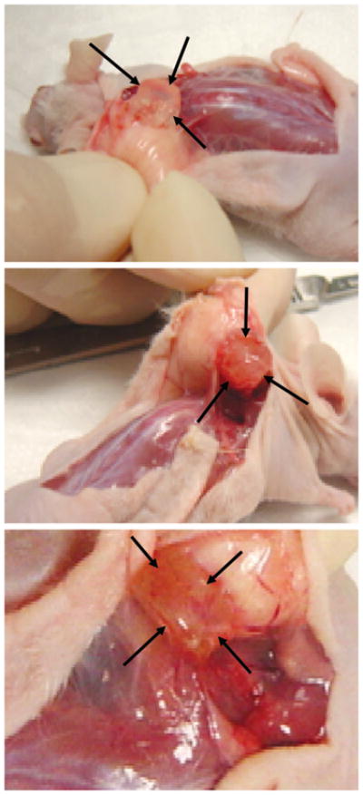
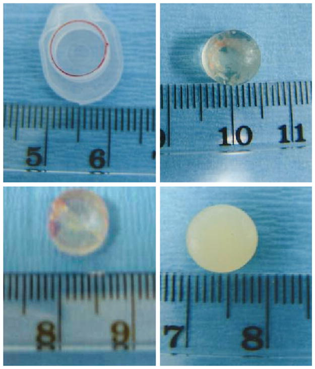
Methods: Human mesenchymal stem cells were isolated from bone marrow of a healthy donor and differentiated into adipogenic cells. Adipocytes derived from these cells were encapsulated in a poly(ethylene glycol)-based hydrogel shaped into a generic cylinder (n = 6 implants), with hydrogel encapsulating human mesenchymal stem cells (n = 6) and cell-free hydrogel (n = 6) as controls. Porous collagen sponges were also used to seed human mesenchymal stem cell-derived adipocytes (n = 6), human mesenchymal stem cells (n = 4), or without cells (n = 4). All poly(ethylene glycol) and collagen constructs were implanted subcutaneously in athymic mice for 4 weeks.

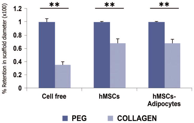
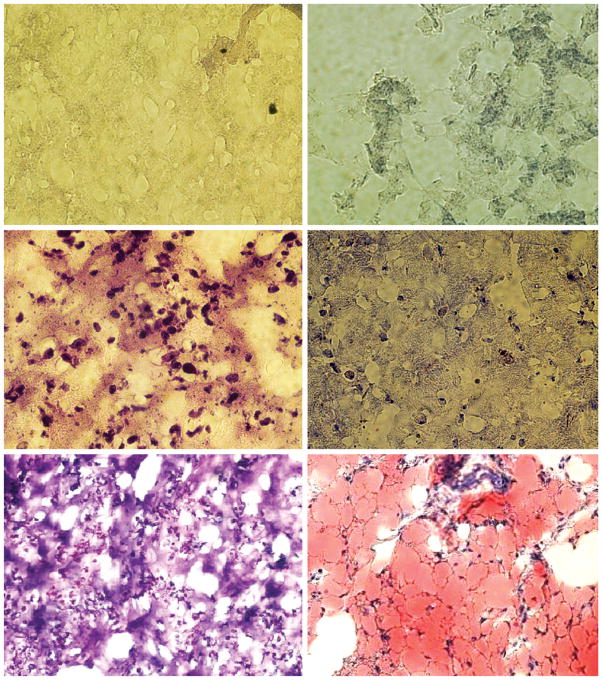
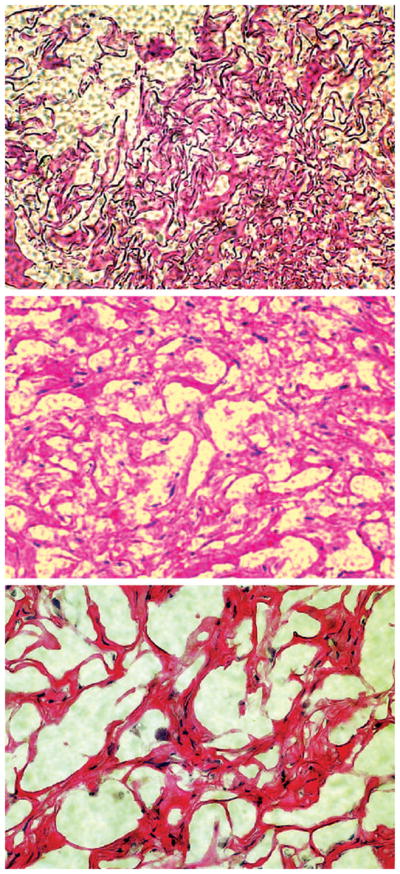
Results: In vivo grafts demonstrated the formation of substantial adipose tissue encapsulating human mesenchymal stem cell-derived adipogenic cells in either poly(ethylene glycol)-based hydrogel or collagen sponge and a lack of adipose tissue formation in cell-free or human mesenchymal stem cell-derived grafts. Engineered adipose tissue in poly(ethylene glycol)-based hydrogel maintained approximately 100 percent of the original dimensions after 4-week in vivo implantation, significantly higher than the approximately 35 to 65 percent volume retention by collagen sponge.

Conclusions: These findings demonstrate that the predefined shape and dimensions of adipose tissue engineered from human mesenchymal stem cells can be maintained after in vivo implantation. These data further indicate the potential for autologous applications in reconstructive and plastic surgery procedures.


Figures

Fig. 1
Fig. 1
Human mesenchymal stem cells and their adipogenic differentiation in monolayer culture. (Above, left) Phase-contrast image of human mesenchymal stem cells undergoing proliferation. Note the fibroblast-like appearance of human mesenchymal stem cells (cf. Alhadlaq and Mao, 2004). (Below, left) Negative Oil-Red O staining of human mesenchymal stem cells without adipogenic induction, as in above, left. (Above, right) After treatment with adipogenic supplements of dexamethasone, insulin, and isobutyl-methylxanthine, the same population of human mesenchymal stem cells as in above, left treated with adipogenic stimulating medium for 1 week showed different cell morphology and the presence of rounded extracellular matrix vacuoles. (Below, right) Positive Oil-Red O staining of lipid vacuoles (arrow) of human mesenchymal stem cells treated with adipogenic induction indicates that human mesenchymal stem cells had differentiated into adipogenic cells.
Fig. 2
Fig. 2
Harvest of adipogenic poly(ethylene glycol)-based hydrogel grafts from human mesenchymal stem cells and control groups after in vivo implantation in athymic mice. (Above) Representative cell-free poly(ethylene glycol)-based hydrogel construct (between arrows) showing retention of the original size (9-mm diameter). (Center) Representative poly(ethylene glycol)-based hydrogel construct encapsulating human mesenchymal stem cells without adipogenic differentiation (between arrows) showing that poly(ethyleneglycol)-based hydrogel adhered to surrounding host tissue and retained the original size (9-mm diameter). (Below) Representative poly(ethylene glycol)-based hydrogel construct (between arrows) encapsulating adipogenic cells derived from human mesenchymal stem cells showing its adhesion to surrounding host tissue and retention of the original size (9-mm diameter; greater magnification).
Fig. 3
Fig. 3
Shape, dimensions, and photo-opaqueness of in vivo harvested poly(ethylene glycol)-based hydrogel construct encapsulating engineered adipose tissue from human mesenchymal stem cells and control groups. (Above, left) Plastic cap of a 1.5-ml microcentrifuge tube (9-mm diameter) used as a generic mold of the shape and dimensions for engineered adipose tissue. (Above, right) Harvested cell-free poly(ethylene glycol)-based hydrogel is largely transparent. (Below, left) Poly(ethylene glycol)-based hydrogel encapsulating human mesenchymal stem cells (without adipogenic differentiation) showing some photo-opacity. (Below, right) Poly(ethylene glycol)-based hydrogel encapsulating adipogenic cells derived from human mesenchymal stem cells showing substantial photo-opacity. All poly(ethylene glycol) grafts maintained the original shape and dimensions (cf. above, left).
Fig. 4
Fig. 4
Quantitative analysis of retention of scaffold diameters (×100 percent ± SD) of harvested poly(ethylene glycol) and collagen grafts after 4 weeks of in vivo implantation (hMSCs, human mesenchymal stem cells). Dark blue bar graphs represent poly(ethylene glycol)-based hydrogel grafts; the lighter bar graphs represent collagen grafts. Poly(ethylene glycol)-based hydrogel without cells (n = 6), encapsulating human mesenchymal stem cells (n = 6), or encapsulating human mesenchymal stem cell– derived adipocytes(n = 6)maintained the original diameter of the poly(ethyleneglycol) scaffold nearly 100 percent. In comparison, collagen sponges without seeded cells (n = 4) lost the original diameter by 65.0 ± 5.0 percent, whereas collagen sponges seeded with human mesenchymal stem cells (n = 4) and with human mesenchymal stem cell– derived adipocytes (n = 6) lost the original diameters by 31.7 ± 5.8 percent and 31.7 ± 5.4 percent, respectively. **p < 0.01 (analysis of variance with Bonferroni). Analysis of variance was used for both within- and between-group comparisons, although p < 0.01 is only shown for within-group comparisons.
Fig. 5
Fig. 5
Representative hematoxylin and eosin and Oil-Red O staining of tissue-engineered poly(ethylene glycol)-based hydrogel grafts retrieved after 4-week in vivo implantation in the dorsum of athymic mice (hMSCs, human mesenchymal stem cells). (Above) Representative hematoxylin and eosin– and Oil-Red O–stained micrographs of cell-free control poly(ethylene glycol)-based hydrogels construct showing neither resident cells nor lipid vacuoles. (Center, left) Representative hematoxylin and eosin–stained micrograph of poly(ethylene glycol)-based hydrogel construct encapsulating human mesenchymal stem cells demonstrates abundant resident cells. (Center, right) Representative Oil-Red O–stained micrograph of poly(ethylene glycol)-based hydrogel construct encapsulating human mesenchymal stem cells demonstrates a lack of lipid vacuoles. (Below, left) Representative hematoxylin and eosin–stained micrograph of poly(ethylene glycol)-based hydrogel construct encapsulating human mesenchymal stem cell– derived adipogenic cells demonstrates abundant resident cells among irregular islands of space. (Below, right) Representative Oil-Red O–stained micrograph of poly-(ethylene glycol)-based hydrogel construct encapsulating human mesenchymal stem cell– derived adipogenic cells demonstrates abundant lipid vacuoles among flattened cells that resemble adipocytes. It is probable that lipid vacuoles occupied the irregular islands of space seen in below, left (original magnification, ×10).
Fig. 6
Fig. 6
Representative hematoxylin and eosin and Oil-Red O staining of tissue-engineered collagen sponge grafts retrieved after 4-week in vivo implantation in the dorsum of athymic mice (hMSCs, human mesenchymal stem cells). (Above and center) Representative hematoxylin and eosin– and Oil-Red O–stained micrographs of cell-free control collagen sponge construct showing the presence of resident cells among apparent collagen bundles. (Below) Representative hematoxylin and eosin–stained micrograph of collagen sponge construct seeded with human mesenchymal stem cells demonstrates resident cells among apparent collagen bundles.

References

    1. Patrick CW, Jr, Chauvin PB, Reece GP. Preadipocyte seeded PLGA scaffolds for adipose tissue engineering. Tissue Eng. 1999;5:139. - PubMed
    1. Beahm EK, Walton RL, Patrick CW., Jr Progress in adipose tissue construct development. Clin Plast Surg. 2003;30:547. - PubMed
    1. Alster TS, West TB. Human-derived and new synthetic injectable materials for soft-tissue augmentation: Current status and role in cosmetic surgery. Plast Reconstr Surg. 2000;105:2515. - PubMed
    1. American Society of Plastic Surgeons (Web site) [Accessed March 26, 2005.]. Available at: http//www.plasticsurgery.org.
    1. Katz AJ, Llull R, Hedrick MH, Futrell JW. Emerging approaches to the tissue engineering of fat. Clin Plast Surg. 1999;26:587. - PubMed

Publication types