. 2007 Feb;117(2):375-86.
doi: 10.1172/JCI29709. Epub 2007 Jan 25.

Adiponectin modulates inflammatory reactions via calreticulin receptor-dependent clearance of early apoptotic bodies

Affiliations

Adiponectin modulates inflammatory reactions via calreticulin receptor-dependent clearance of early apoptotic bodies

Yukihiro Takemura et al. J Clin Invest. 2007 Feb.

Abstract

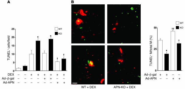
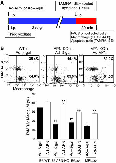
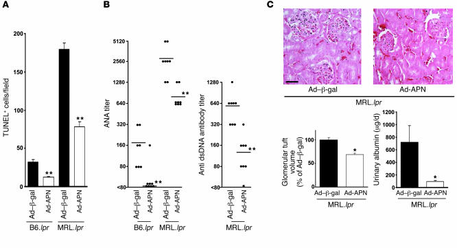
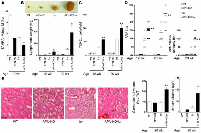
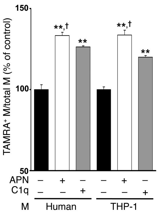
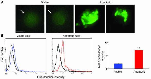
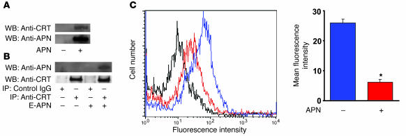
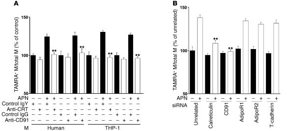
Obesity and type 2 diabetes are associated with chronic inflammation. Adiponectin is an adipocyte-derived hormone with antidiabetic and antiinflammatory actions. Here, we demonstrate what we believe to be a previously undocumented activity of adiponectin, facilitating the uptake of early apoptotic cells by macrophages, an essential feature of immune system function. Adiponectin-deficient (APN-KO) mice were impaired in their ability to clear apoptotic thymocytes in response to dexamethasone treatment, and these animals displayed a reduced ability to clear early apoptotic cells that were injected into their intraperitoneal cavities. Conversely, adiponectin administration promoted the clearance of apoptotic cells by macrophages in both APN-KO and wild-type mice. Adiponectin overexpression also promoted apoptotic cell clearance and reduced features of autoimmunity in lpr mice whereas adiponectin deficiency in lpr mice led to a further reduction in apoptotic cell clearance, which was accompanied by exacerbated systemic inflammation. Adiponectin was capable of opsonizing apoptotic cells, and phagocytosis of cell corpses was mediated by the binding of adiponectin to calreticulin on the macrophage cell surface. We propose that adiponectin protects the organism from systemic inflammation by promoting the clearance of early apoptotic cells by macrophages through a receptor-dependent pathway involving calreticulin.


Figures

Figure 1
Figure 1. APN deficiency leads to the accumulation of apoptotic debris.
(A) WT and APN-KO mice were treated with dexamethasone (DEX), and thymi were stained for apoptotic cells with TUNEL. In some experiments, mice received an i.v. infusion of Ad-APN or Ad–β-gal 2 days prior to injection of dexamethasone. The number of TUNEL-positive cells per microscopic field for the different experimental conditions is reported. *P < 0.05 versus WT; P < 0.05 versus Ad–β-gal for WT or APN-KO (n = 3–6). (B) Adiponectin stimulates macrophage engulfment of TUNEL-positive apoptotic debris in thymi of dexamethasone-treated mice. Histological sections were stained with TUNEL (green) and anti-CD11b antibody (red). Scale bar: 20 μm. Colocalization is indicated by yellow in the merged images. ††P < 0.01 versus Ad–β-gal for WT or APN-KO (n = 3–6). M, macrophage.
Figure 2
Figure 2. Systemic delivery of adiponectin promotes the uptake of apoptotic debris by peritoneal macrophages.
(A) Strains of mice were injected with Ad–β-gal or Ad-APN on the same day as thioglycollate treatment. Circulating adiponectin levels at the time of sacrifice were 12.5 ± 1.6 μg/ml in B6.lpr/Ad–β-gal and 19.5 ± 2.4 μg/ml in B6.lpr/Ad-APN. TAMRA, SE–labeled apoptotic Jurkat T cells were injected into the peritoneum of the indicated strains of mice 3 days after the administration of thioglycollate. After 30 minutes, peritoneal cells were removed by lavage and subjected to flow cytometry. (B) Phagocytosis was scored as the percentage of F4/80-positive macrophages that also stained positive for TAMRA, SE. Scatter plots show representative flow cytometry data for 3 experimental conditions. Dual-labeled cells are represented in the upper right quadrant. **P < 0.01 versus Ad–β-gal; ††P < 0.01 versus WT (n = 6).
Figure 3
Figure 3. Systemic delivery of adiponectin decreases remnant apoptotic levels and inflammation in lpr strains of mice.
(A) Adenovirus-mediated overexpression of adiponectin decreases apoptotic cells in lymph nodes of B6.lpr mice at the age of 12 weeks and MRL.lpr mice at the age of 20 weeks. Lymph node sections were stained with TUNEL and quantified by microscopy (n = 6–8). (B) ANA titers and anti-dsDNA antibody titer in the serum of B6.lpr or MRL.lpr mice at 14 days after Ad-APN or Ad–β-gal administration. **P < 0.01 versus Ad–β-gal. (C) Adiponectin overexpression influences renal function and morphology. Photographs show representative glomeruli of MRL.lpr mice treated with Ad–β-gal or Ad-APN 14 days prior to sacrifice. Glomerular tuft volume of control is 3.1 × 10⁵ ± 0.1 × 105 μm3. Scale bar: 50 μm. Urinary albumin is reported as μg protein/d. *P < 0.05 versus Ad–β-gal (n = 8).
Figure 4
Figure 4. B6/lpr mice deficient in adiponectin display impaired clearance of apoptotic cells and increased systemic inflammation.
(A) Impaired clearance of apoptotic cells by peritoneal macrophages in APN-KO/lpr mice compared with APN-KO and lpr mice. Experiments were conducted with the indicated strains of mice. Phagocytosis of apoptotic Jurkat cells was assessed by flow cytometric analysis of F4/80- and TAMRA, SE–positive cells as described in the Figure 1C legend. *P < 0.05 versus B6/lpr APN-KO (n = 4–6). (B) APN-KO/lpr mice have larger submandibular lymph nodes compared with lpr mice. Inset shows a representative photograph of submandibular lymph nodes from each strain of mouse at the age of 20 weeks. Scale bar: 5 mm. Submandibular lymph nodes were excised from the indicated strains of mice at 12 or 20 weeks and weighed. *P < 0.05 versus lpr (n = 6–14). (C) Adiponectin deficiency increases the frequency of apoptotic cells in lymph nodes of B6.lpr mice. Mice were sacrificed at 12 or 20 weeks of age, and TUNEL-positive cells in sections of submandibular lymph nodes were assessed. ND, not detectable. **P < 0.01 versus lpr (n = 6–14). (D) Adiponectin deficiency increases autoreactive antibody titer in B6.lpr mice. ANA titers and anti-dsDNA antibody titers in the sera of each strain of mouse were determined at 12 or 20 weeks of age. **P < 0.01 versus lpr (n = 3–14). (E) Adiponectin deficiency promotes kidney disease in B6.lpr mice. Glomerular tuft volume was determined in histological sections of kidney from the indicated strains of 20-week-old mice. Photographs show representative glomeruli. The calculated glomerular tuft volume of control is 1.1 × 10⁵ ± 0.1 × 105 μm3. Scale bar: 50 μm. **P < 0.01 versus lpr. The daily excretion of urinary albumin was determined immediately prior to sacrifice. *P < 0.05 versus lpr (n = 4–10).
Figure 5
Figure 5. Adiponectin promotes the phagocytosis of apoptotic bodies by macrophages in vitro.
Apoptotic Jurkat T cells were preincubated for 1 hour with recombinant adiponectin from baculovirus-insect (APN) (50 μg/ml), human C1q (50 μg/ml), or vehicle. Macrophages were then incubated for 30 minutes with TAMRA, SE–labeled Jurkat cells that were either viable or apoptotic due to UVB exposure. Upon mixing apoptotic cells with macrophages, adiponectin and C1q were diluted to a final concentration of 10 μg/ml. Phagocytosis was assessed by flow cytometry. Macrophages were stained with FITC-conjugated anti-human macrophage antibody, and the percentage of phagocytic macrophages was calculated as TAMRA, SE–positive (+) macrophages/total macrophages × 100%. Phagocytic macrophages of control were 32.8% ± 1.0% (human) and 23.6% ± 0.4% (THP-1). **P < 0.01 versus vehicle; P < 0.05 versus C1q (n = 6–7).
Figure 6
Figure 6. Apoptosis increases the binding of adiponectin to Jurkat T cells.
(A) Fluorescence microscopy of FITC-APN to viable (left panel) and apoptotic (right panel) Jurkat T cells. Binding to viable cells was diffuse whereas binding to apoptotic cells was intense and uneven within individual cells. Scale bar: 5 μm. (B) Left panels: representative flow cytometric analyses of FITC-APN to viable (blue) and apoptotic Jurkat T (red) cells. Control (black) represents cells incubated with FITC-conjugated human serum albumin. Right panel: Quantitation of FITC-APN binding to viable and apoptotic cells. **P < 0.01 versus viable cells (n = 3).
Figure 7
Figure 7. Adiponectin interacts with calreticulin on the macrophage cell surface.
(A) Calreticulin is immunoprecipitated by histidine-tagged adiponectin (APN) from detergent-solubilized THP-1 membranes. Membrane fractions were incubated in the presence or absence of polyhistidine-APN and then precipitated with nickel resin. The resin was then treated with a molar excess of histidine to release precipitated proteins, and this material was subjected to SDS-PAGE followed by immunoblot analysis with anti-calreticulin (anti-CRT) and anti-adiponectin (anti-APN) antibodies. WB, Western blot. (B) Adiponectin prepared from E. coli (E-APN) was immunoprecipitated from detergent-solubilized THP-1 membranes by anti-calreticulin antibodies. THP-1 membrane fractions were incubated in the presence or absence of polyhistidine APN and then subjected to immunoprecipitation with anti-CRT or control IgG. Immunoprecipitated material was then subjected to SDS-PAGE, and Western blot analysis was performed with anti-APN or anti-CRT antibodies. (C) Adiponectin inhibits the binding of anti-calreticulin antibody to macrophages. THP-1 macrophages were preincubated with 200 μg/ml adiponectin (red) or vehicle (blue) for 60 minutes followed by incubation with anti-calreticulin antibody (10 μg/ml) for 60 minutes. Cells incubated with chicken IgY followed by treatment with FITC-conjugated secondary antibody served as control. Cells were then incubated with FITC-conjugated secondary antibody to anti-calreticulin antibody and analyzed by flow cytometry. *P < 0.05 versus vehicle (n = 3).
Figure 8
Figure 8. Calreticulin and CD91 are essential for adiponectin-stimulated uptake of apoptotic cells.
(A) Anti-calreticulin antibody and anti-CD91 antibody inhibit adiponectin-stimulated apoptotic cell phagocytosis by macrophages. Each type of macrophage was preincubated with anti-CRT antibody or control chicken IgY or with anti-CD91 antibody or control IgG for 60 minutes. TAMRA, SE–labeled apoptotic cells were preincubated with recombinant adiponectin or vehicle, and uptake of apoptotic debris was determined by flow cytometric analysis. Data are expressed relative to control from human and THP-1 monocytes. Control human and THP-1 macrophages (anti-human macrophage antibody-positive) were 34.1% ± 0.8% and 24.9% ± 1.1% dual-positive for TAMRA, SE, respectively. **P < 0.01 versus IgY or IgG (n = 6–7). (B) Adiponectin-stimulated apoptotic cell phagocytosis by macrophages was inhibited by downregulation of calreticulin or CD91 with siRNA but not siRNA targeting the putative adiponectin receptors. The in vitro phagocytosis assay analyzed dual-positive by cells flow cytometry. Control THP-1 macrophages were 24.3% ± 1.0% positive for TAMRA, SE. **P < 0.01 versus unrelated siRNA with adiponectin (n = 6–7).

References

    1. Weisberg S.P., et al. Obesity is associated with macrophage accumulation in adipose tissue. J. Clin. Invest. 2003;112:1796–1808. doi: 10.1172/JCI200319246. - DOI - PMC - PubMed
    1. Ruan H., Lodish H.F. Regulation of insulin sensitivity by adipose tissue-derived hormones and inflammatory cytokines. Curr. Opin. Lipidol. 2004;15:297–302. - PubMed
    1. Spiegelman B.M., Choy L., Hotamisligil G.S., Graves R.A., Tontonoz P. Regulation of adipocyte gene expression in differentiation and syndromes of obesity/diabetes. J. Biol. Chem. 1993;268:6823–6826. - PubMed
    1. Maeda K., et al. cDNA cloning and expression of a novel adipose specific collagen-like factor, apM1 (AdiPose Most abundant Gene transcript 1). Biochem. Biophys. Res. Commun. 1996;221:286–289. - PubMed
    1. Hu E., Liang P., Spiegelman B.M.1996. . AdipoQ is a novel adipose-specific gene dysregulated in obesity. J. Biol. Chem. 27110697–10703. - PubMed

Publication types

MeSH terms